This was a 57 year old gentleman , with complaints of difficulty in passing urine and dysuria of 1 month duration.
His upper abdominal scan was normal . His left kidney was normal.
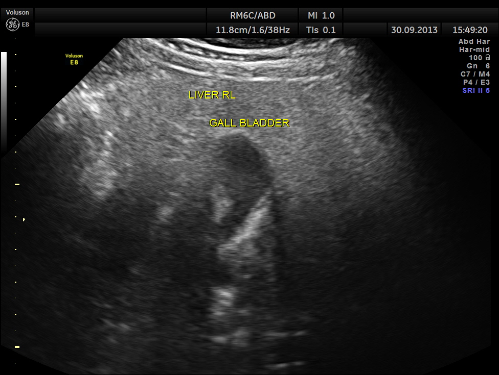
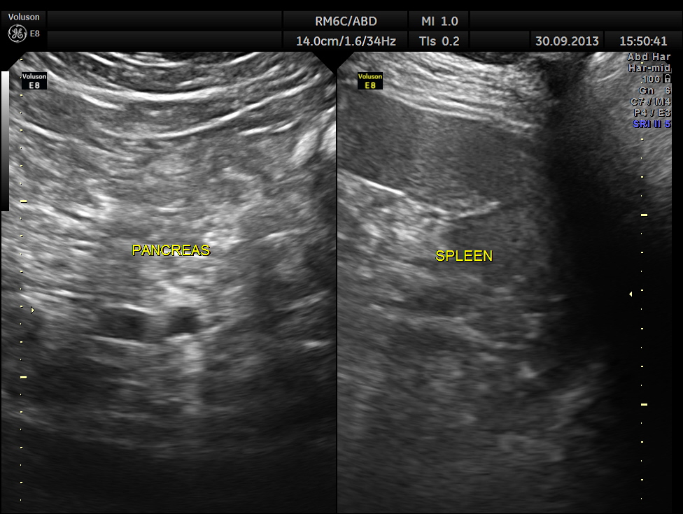
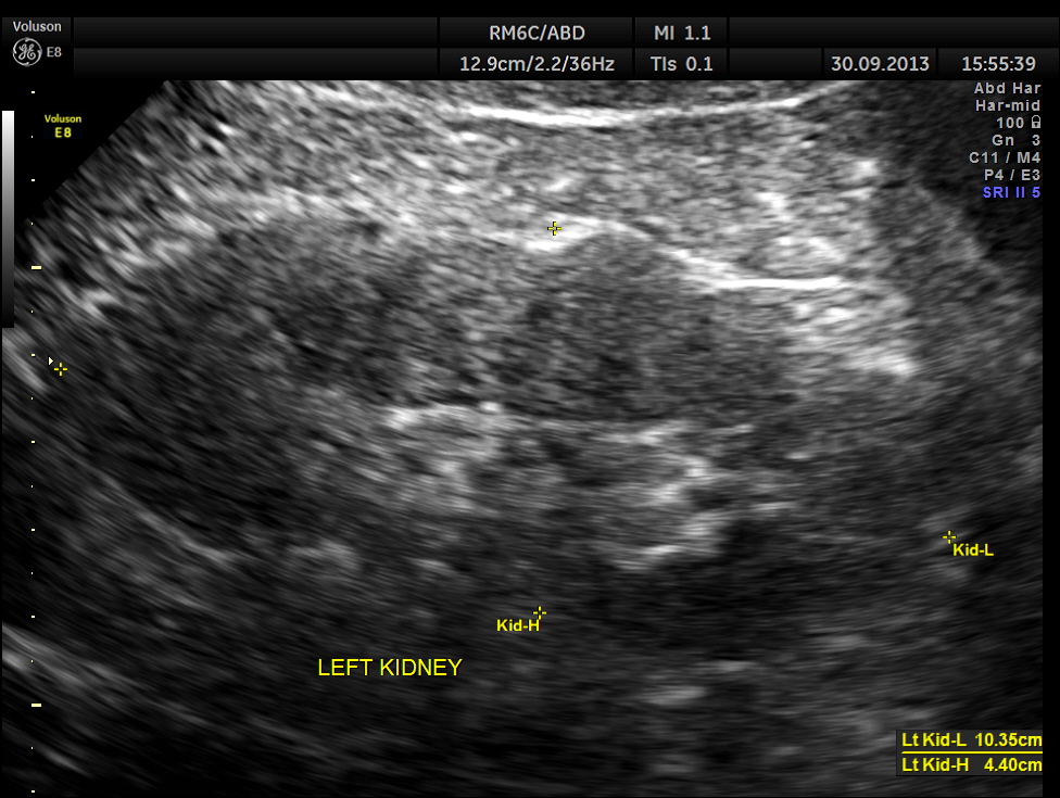
the right kidney showed moderate to severe hydro-nephrosis


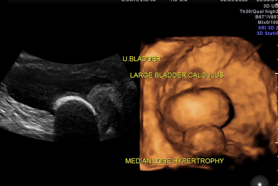
urinary bladder showed a large calculus and prominent swelling of the median lobe of the prostate.
the following images show the 2 d and 3 d appearance of the same.
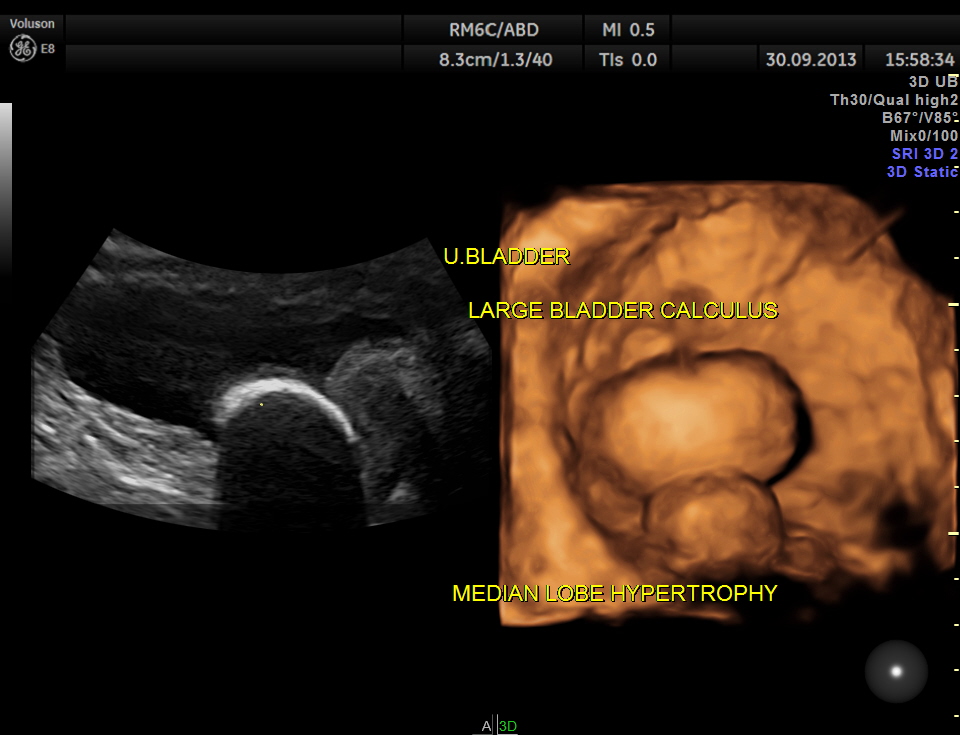
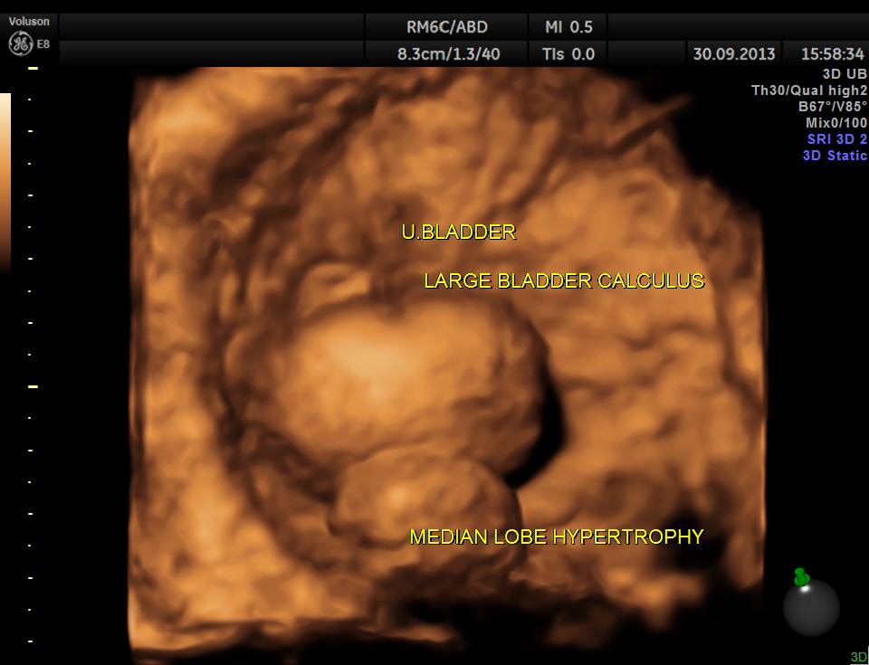
enlarged prostate and median lobe hypertrophy
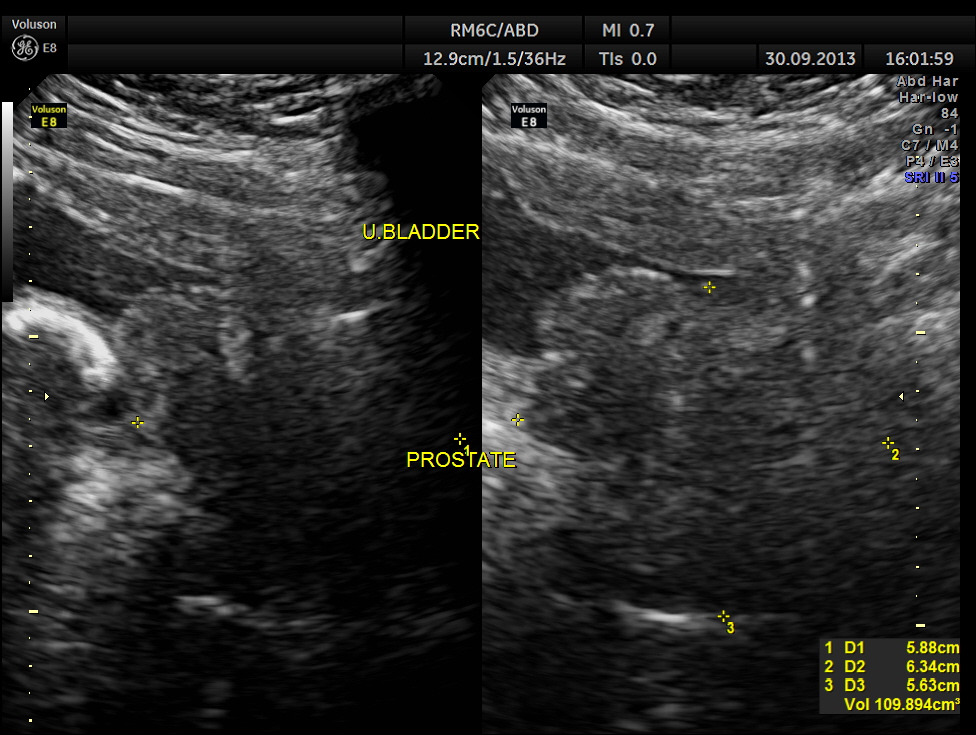
2 D and 3 D of the bladder calculus alone.
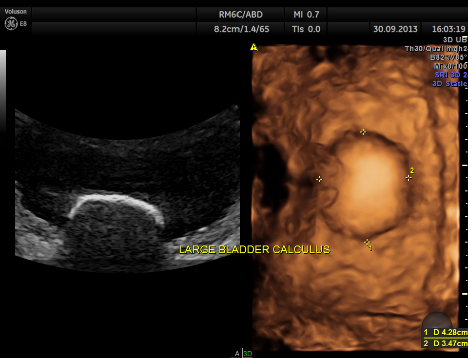
multi- planar view of the bladder calculus
r 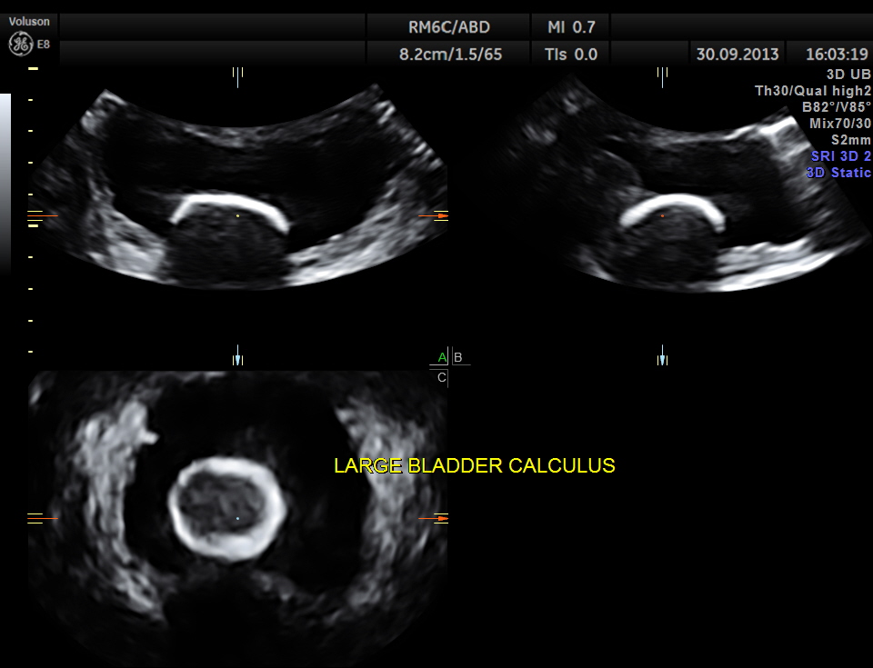
2 D and 3 D views of the median lobe hypertrophy
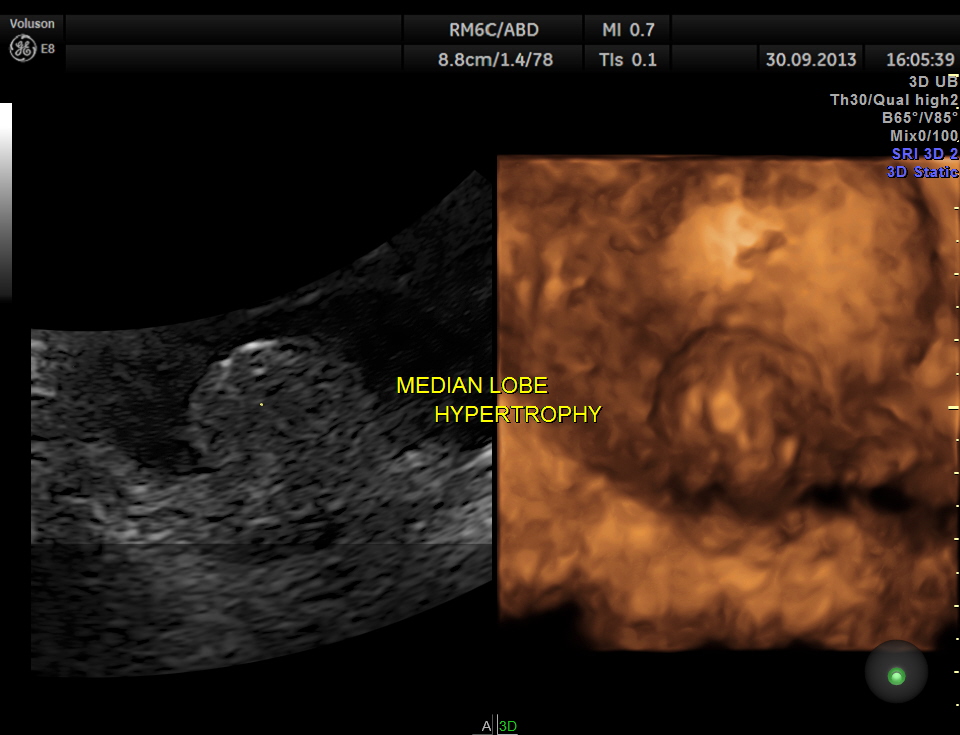
the median lobe hypertrophy in 2 D
The diagnosis given was Large calculus in the urinary bladder , Severe prostatic enlargement with prominent median lobe hypertrophy , causing Right sided obstructive uropathy.
The diagnosis was made with the 2D images , but the 3 D images were very helpful in explaining to the patient.


Life like pictures clearly indicative,very much appreciate this posting.
LikeLike
Thanks sir
LikeLike
Very clear 3D pictures .impressed about your interest & dedication.god bless you
LikeLike
Thank you
LikeLike
As usual, very illustrative pictures. Thanks for showing us the great variety of patients that you see! What have they offered this patient in terms of treatment?
LikeLike
About the treatment plans, can I get back to you some time later
Thanks for your comments
LikeLike
very impressive.
i would like to know how i could have a better understanding of 3d and 4d
thanks
LikeLike
Well done sir.I admire your efforts & experties.
LikeLike
Thanks sir
LikeLike
Pingback: Acute Bilateral Obstructive Uropathy | Find Me A Cure
according to cause
LikeLike