
This was a 39 year old lady presenting with abnormal uterine bleeding for the past 1 month .
The trans abdominal images are given first. A cervical polyp was seen .


- coronal view ( trans abdominal )
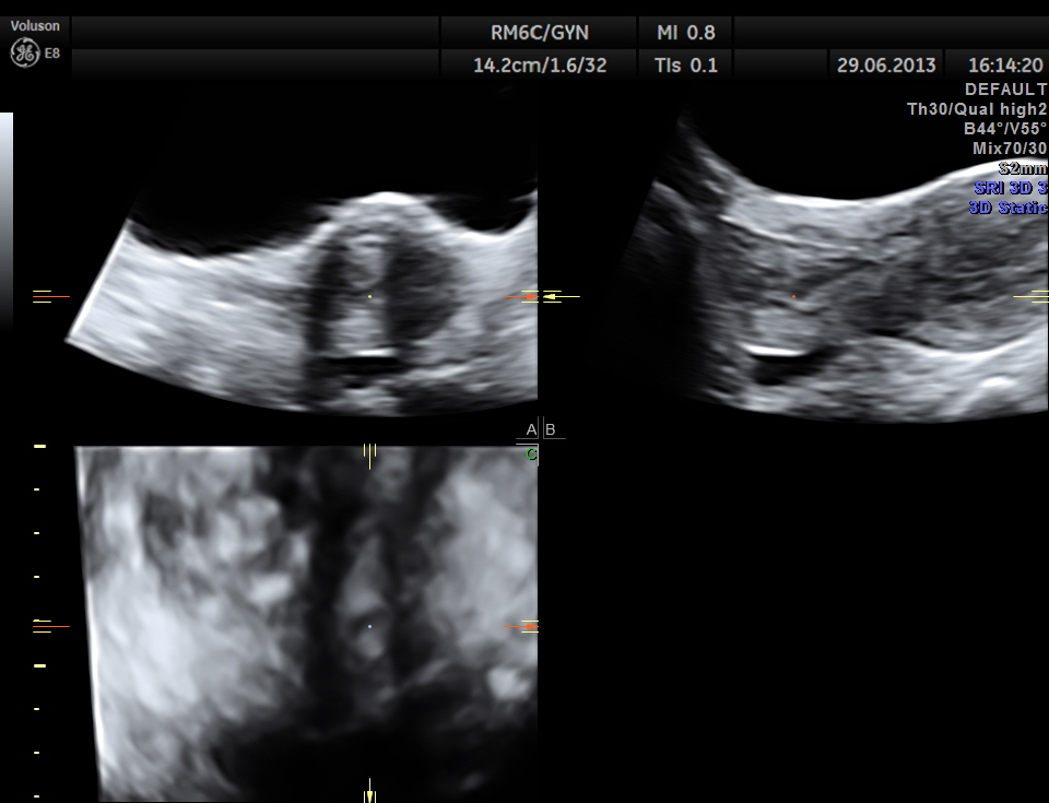
trans abdominal multi planar view

trans abdominal colour doppler showing a long vascular stalk
the following images are acquired trans vaginally and the starking difference in clarity can be appreciated.
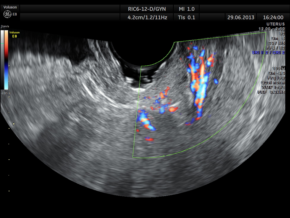
the increased vascularity of the polyp in the cervix can be seen

the multi planar 3 d image shows increased flow in the polyp and the stalk arising from the endometrium

the vascular stalk is seen

reconstructed image showing the stalk arising from endometrium and the polyp in the cervix
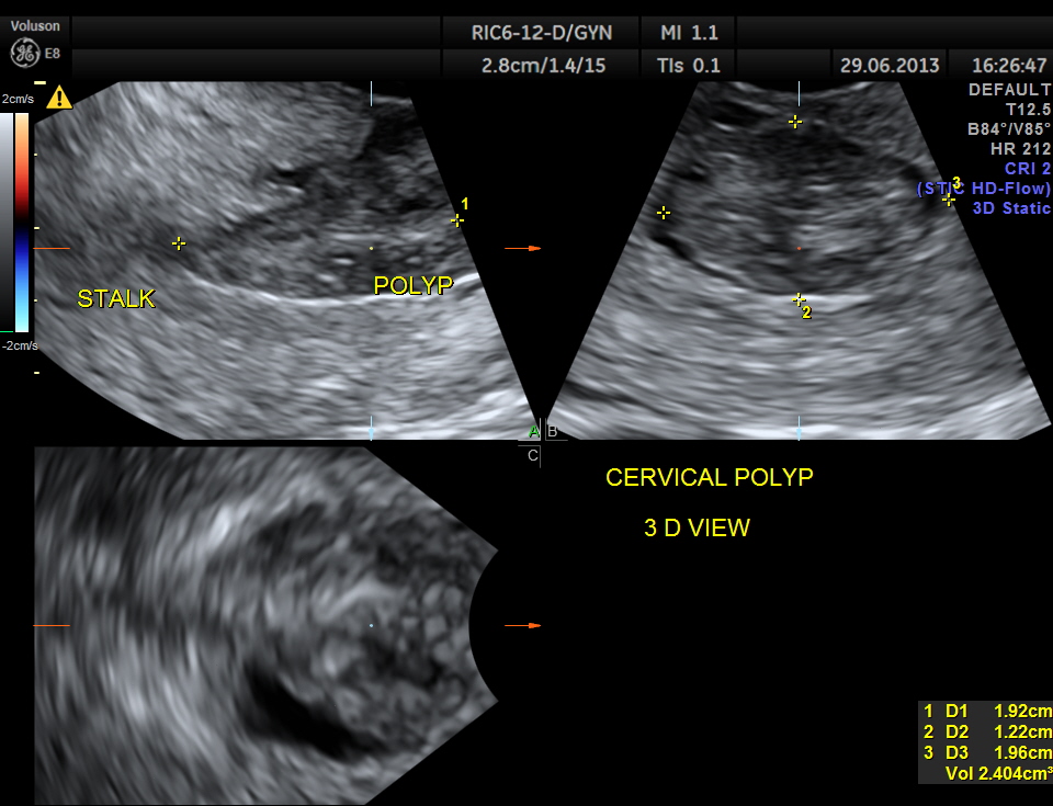
multi planar image showing the polyp
The diagnosis offered was an endometrial polyp with a long stalk situated in the cervix.
The gynaecologist did a polypectomy and cervical and endometrial biopsy.
The histopathology report was : Endometrial polyp ; Chronic cervicitis .
This case illustrates the superiority of trans vaginal scan over the trans abdominal mode ( though the diagnosis could have been made trans abdominally also.)

Pingback: ENDOMETRIAL POLYP WITH A STALK PRESENTING IN THE CERVIX | When I turned 53
Great job!
LikeLike
Awesome !
LikeLike
A remarkable representation. The 3D image is fascinating.
LikeLike
Thanks
LikeLike