This was a 40-year-old lady who presented with acute upper abdominal pain radiating to the back.
Gall stones were seen .
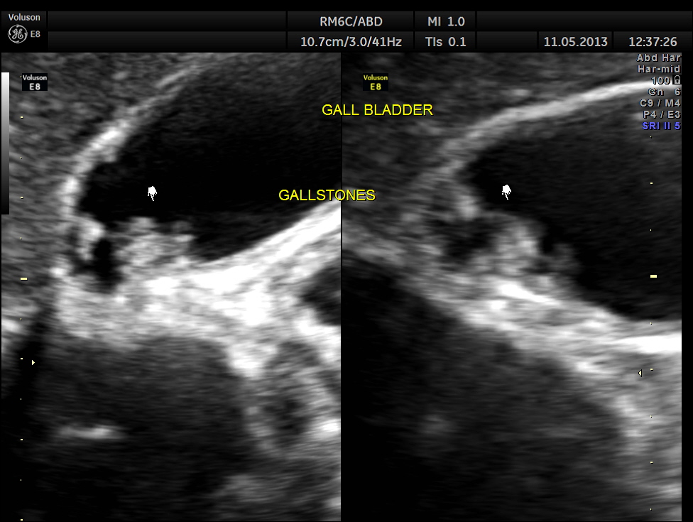

3 d of gallbladder with gallstones
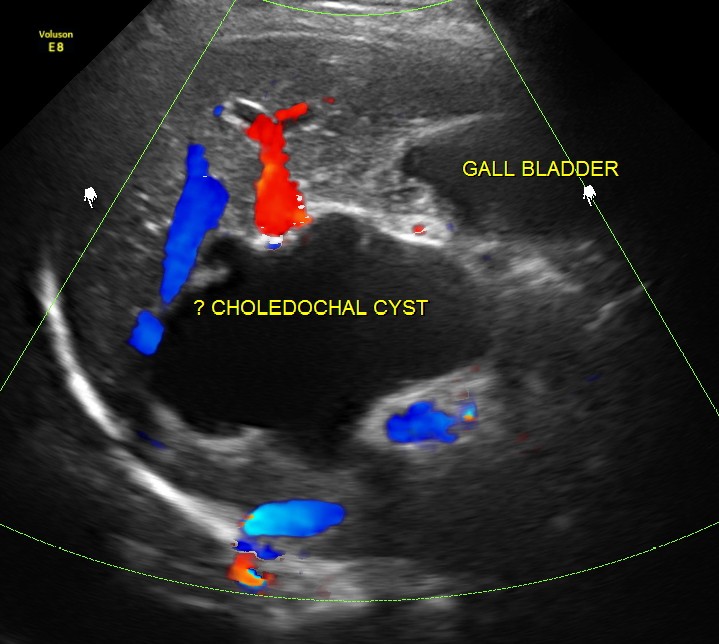
Prominent irregular cysts were seen in both the lobes of the liver ; the cysts were seen to arise from the biliary tract.


Pelvic scan revealed bi cornuate uterus ( incidental ) . She dad 3 children and never had any gynecological complaints

The diagnosis offered was Cholelithiasis , Choledochal cysts Type IV A and incidental bi cornuate uterus.
This patient was further evaluated with MRI , which confirmed the ultrasound findings . She underwent surgery successfully.
The following reference is from wikipedia http://en.wikipedia.org/wiki/Choledochal_cysts
Choledochal cysts are congenital conditions involving cystic dilatation of bile ducts.[1] They are uncommon in western countries[2] but not as rare in East Asian nations like Japan and China.
Most of them present in 1st year of life; adult presentation is rare and usually at this stage is associated with complication . Classic triad ofintermittent abdominal pain, jaundice, and a right upper quadrant abdominal mass is found only in minority of patients.
They were classified into 5 types by Todani in 1977.[3]
Classification was based on site of the cyst or dilatation. Type I to IV has been subtyped.
- Type I: Most common variety (80-90%) involving saccular or fusiform dilatation of a portion or entire common bile duct (CBD) with normalintrahepatic duct.
- Type II: Isolated diverticulum protruding from the CBD.
- Type III or Choledochocele: Arise from dilatation of duodenal portion of CBD or where pancreatic duct meets.
- Type IVa: Characterized by multiple dilatations of the intrahepatic and extrahepatic biliary tree.
- Type IVb: Multiple dilatations involving only the extrahepatic bile ducts.
- Type V: Cystic dilatation of intra hepatic biliary ducts. Not the same etiology as Caroli’s disease.
Choledochal cysts are treated by surgical excision of the cyst with the formation of a roux-en-Y anastomosis to the biliary duct. Future complications include cholangitis and a 2% risk of malignancy, which may develop in any part of the biliary tree.

excellent case
LikeLike
Thanks
LikeLike
Excellent case:-)
LikeLike
Any relationship between the two anomalies; chledonchal cyst and biconuate uterus. Biconuate uterus was obviously congenital. What about chledonchal cyst in a 40 year old woman?
LikeLike
Krishna, thank you for the continuos education.
LikeLike
Thanks sir
LikeLike