This was a 25 year old primi , without a history of consanguinity , sent for 2nd opinion for suspected dilated ventricles .
The following pictures show dilated lateral ventricles and orbital hypotelorism ( meaning an abnormal decrease in the distance between the two eyes (the eyes appear too close together)). in the first picture. Angulation reveals semi-lobar holoprosencephaly.(occipital horns are seen , but other ventricular landmarks are absent ).


holoprosencephaly

holoprosencephaly
The fetal head was in the upper pole , with the spine posterior in location throughout the study.The spine was studied with great difficulty . This was a flipped image showing a neural tube defect.- lumbar meningo myelocele.
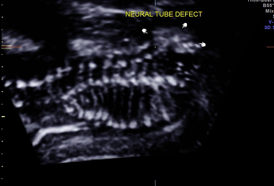
lumbar neural tube defect seen
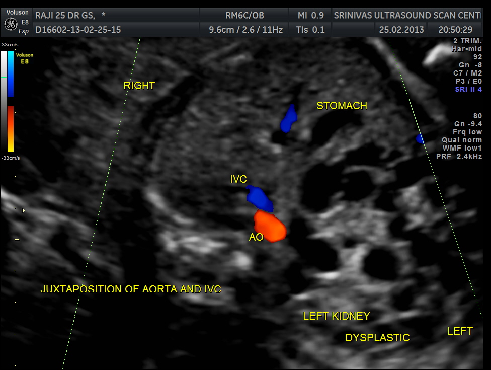
Abdominal transverse view shows aorta and ivc in juxtaposition and on the right side . This is suggestive of heterotaxy – right atrial isomerism. Grossly enlarged dysplastic left kidney is also seen. Right fetal kidney is difficult to visualise in this view. Stomach is in the midline. Heart is on the right side of the chest- dextrocardia
In right atrial isomerism, both atria of the heart are morphological right atria leading to associated abnormalities in the pulmonary venous system. In addition, individuals with right atrial isomerism develop asplenia, a mid-line liver, malrotation of the small intestine and the presence of two morphologic right lungs. Individuals with left atrial isomerism, by comparison, two morphologic left atria, polysplenia, have intestinal malrotation and two morphologic left lungs.

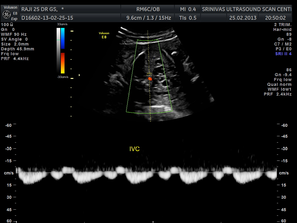
spectral doppler showing aortic flow (above ) and ivc flow ( below image ).
the following colour flow image shows no obvious abnormality.

this shows no obvious abnormality
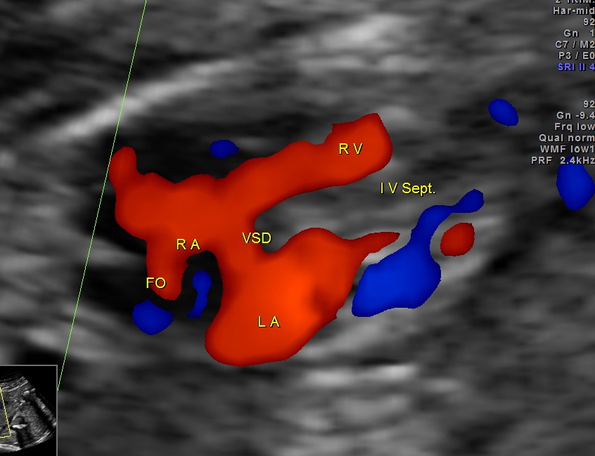
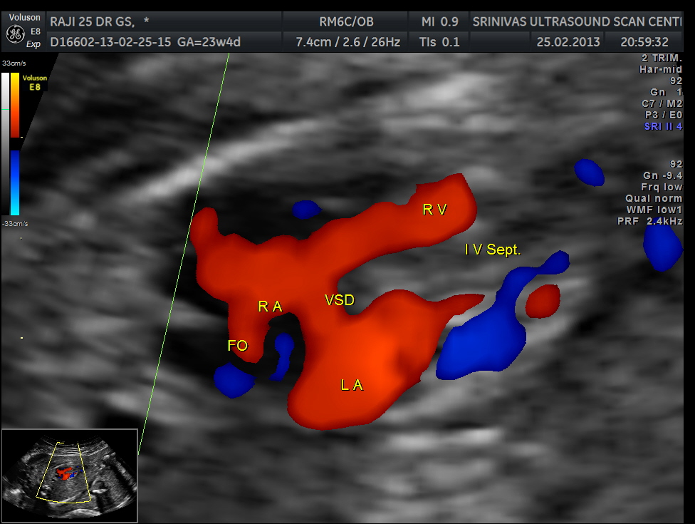
apart from foramen ovale flow , VSD is seen
apart from foramen ovale flow , VSD is seen
Ventricular septal defect seen
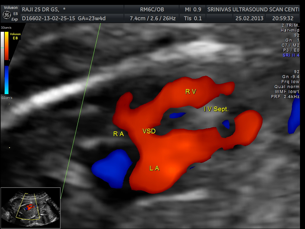
VSD is seen
STIC reconstructed image showing Atrio ventricular valves arising from the same level – feature of heterotaxy / isomerism
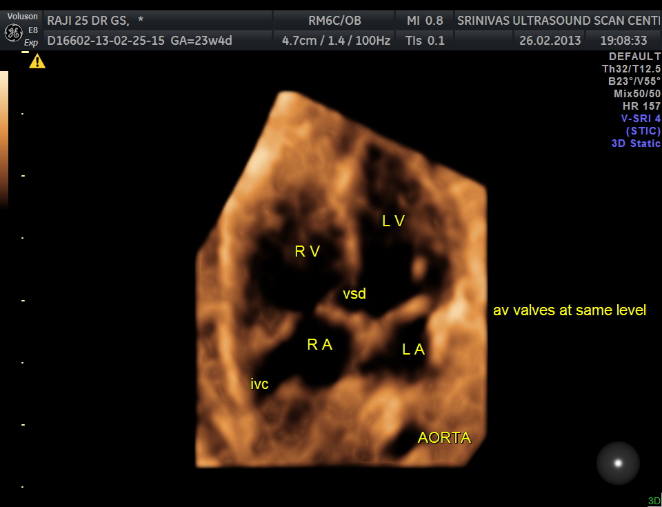
- Both Atrio-ventricular valves at the same level – feature of isomerism
the ventricular size shows an obvious disparity

the left ventricle is obviously smaller than the right ventricle

LV Outflow tact seen
the same picture with the colour flow shows LVOT flow in blue ( away from the transducer ) and the aortic low in red ( towards the transducer ) suggestive of flow reversal .This finding combined with a smaller LV would suggest COARCTATION OF AORTA .(a congenital condition whereby the aorta narrows in the area where the ductus arteriosus(ligamentum arteriosum after regression) inserts.)

reversal of flow in the aorta
The following video shows all of the above cardiac findings .please watch careful.

renal dysplasia – left is grossly enlarged , right is mildly enlarged
Because of the oligohydramnios , visualisation of face , limbs and digits was not good.
So the diagnosis offered was – bilateral renal dysplasia, holoprosencephaly , neural tube defect, complex heart disease ( heterotaxy , vsd ).
Coarctation of aorta was thought of later on examination of the saved images and clips later.

Did the baby have a diaphragmatic hernia also?
LikeLike
No
LikeLike
I don’t agree with the diagnosis of this being a case of holoprosencephaly. Even in mild forms of holoprosencencepahly the frontal horns are fused together. They are not fused in this case. This in all likihood is a case of severe ventriculomegaly (hydrocephalus) and fenestration of the the falx, due to the severe ventriculomegaly.
LikeLike
Discussions and disagreements are a healthy part of a scientific blog . Please read my other posts and give your opinion .
LikeLike
this does not look like holoprosenceply. when there is significant ventriculomegaly, the midline echo may not be seen fully
LikeLike
Sir,
It was a pleasant surprise to see your comment on my blog. The 3rd picture is a 3D picture and the dot is in the midline and shows the connection. I do agree the 1st picture shows only ventriculomegaly.
Thanks for your interaction .
Krishnan
LikeLike
Were you ever able to obtain an etiology for the abnormalities (amniocentesis? microarray? other testing?). We have a similar patient with semilobar holoprosencephaly, heterotaxy, hypoplastic right ventricle, unilateral renal agenesis, also with an absent nose and midline cleft lip and palate. Amniocentesis is normal. Microarray results are pending. Thanks!
LikeLike
Unfortunately the patient was lost for follow up; please share the micro array results when available
LikeLike
We found a case of holoprosencephaly in INM who survived eleven weeks.
LikeLike