This 23 year old primi gravida, with no history of consanguinity was referred for 2nd opinion for evaluation of cystic swelling of the neck in the first trimester.
The following are the 2 d pictures.
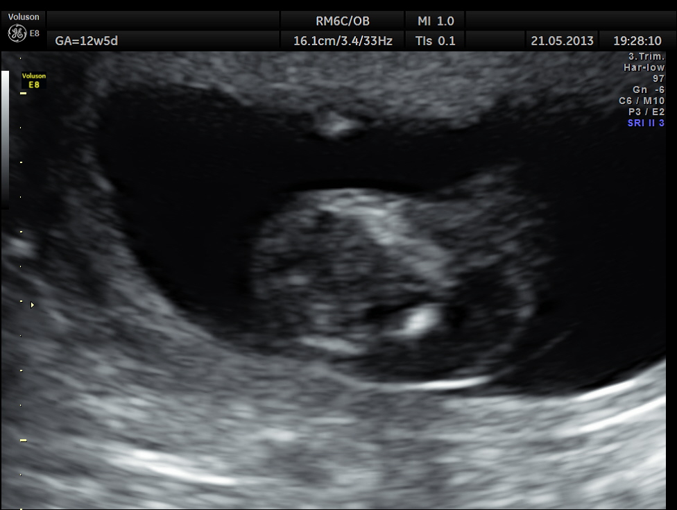
cystic swelling in the posterior aspect of the neck

thin septations seen in the cystic swelling
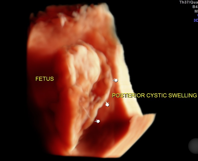
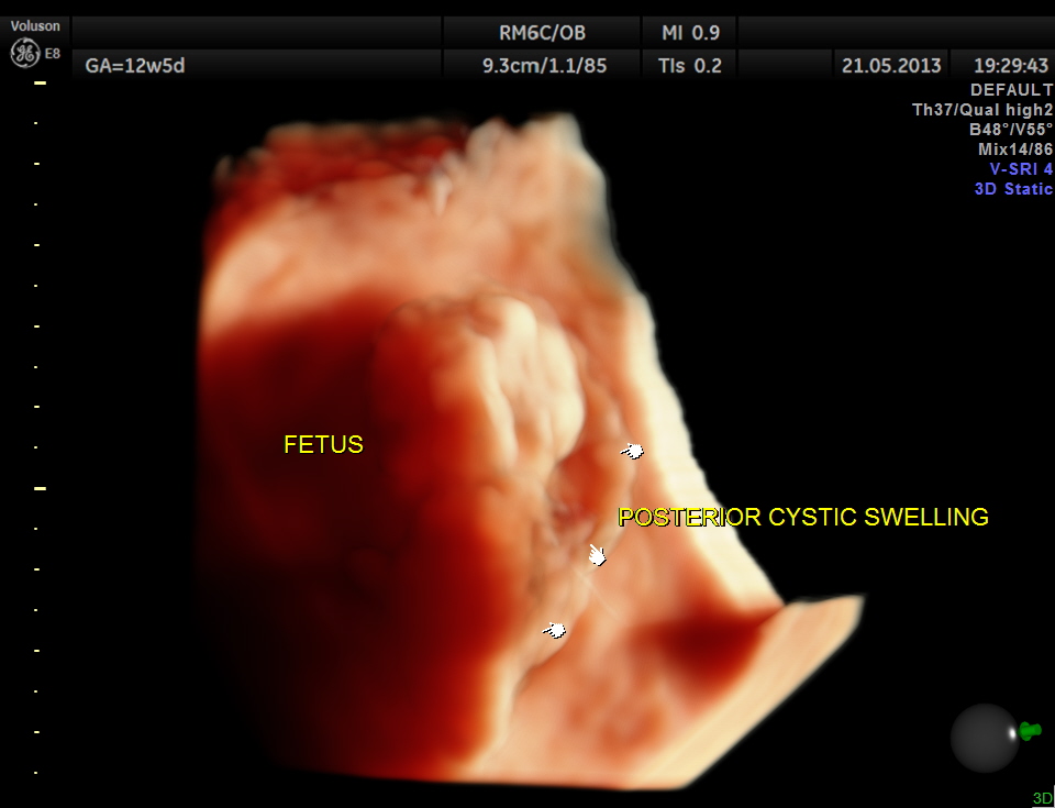
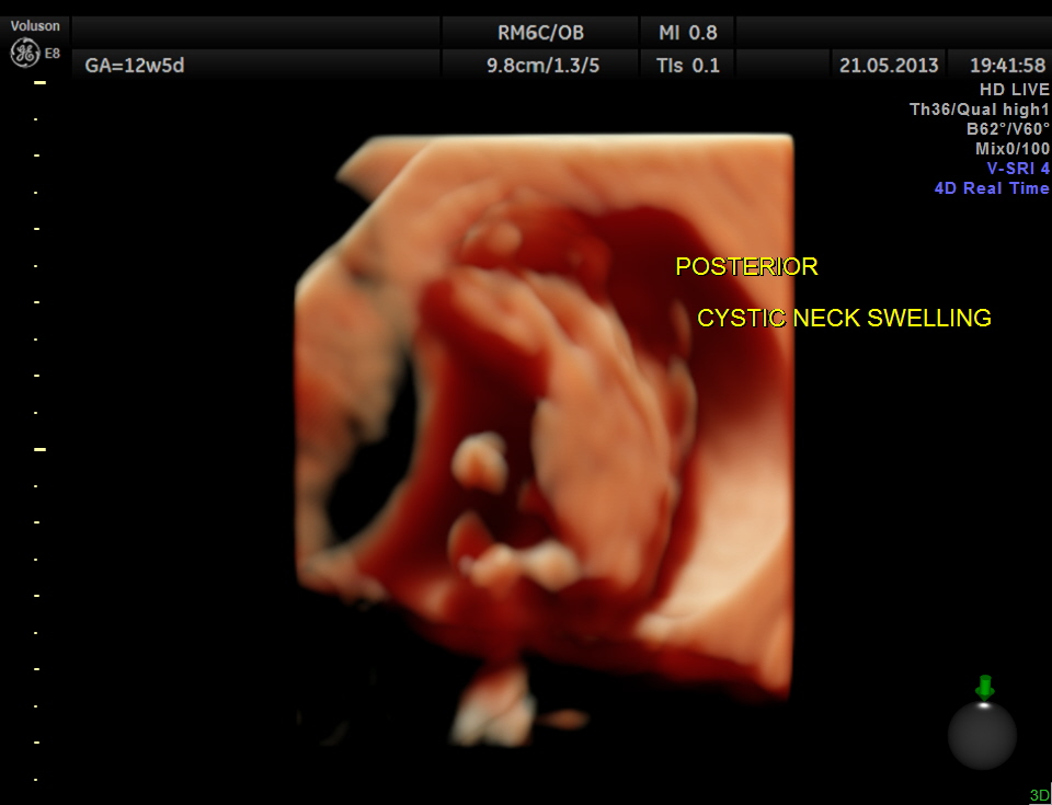
The following is 3D multi dimensional picture.
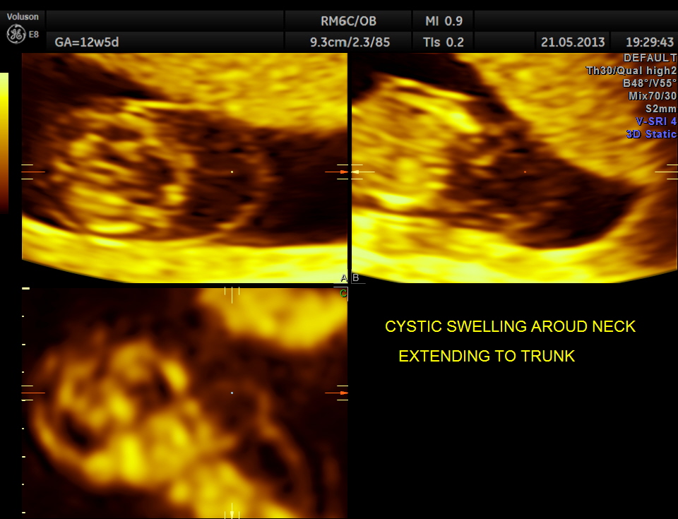
cystic swelling in the posterior aspect of the neck
the following are some reconstructed images.

the following are high definition live images.

The diagnosis offered was cystic hygroma .
The following link is worthwhile exploring.
1992-12-07-21 Cystic hygroma, colli © Varma www.thefetus.net/
http://www.sonoworld.com/Client/TheFetus/page.aspx?id=202
the following is from the above link.
Cystic hygroma colli probably represent the most common cause for a neck mass detected prenatally. Other neck masses detectable ultrasonically include cervical meningomyelocele, hemangioma, teratoma, goiter, sarcoma, and metastatic adenopathy. Occasionally, a large cephalocele may mimic a neck mass. In most of these rare cases, polyhydramnios, hydrops fetalis, or other clinical features signal a careful search of the fetal neck.
Is an abnormal neck mass present?
If so:
1. Is the mass unilateral or bilateral, posterior or anterior ?
2. Is the mass in the midline or not ?
3. What are its echotexture and Doppler characteristics ?
* most bilateral posterior masses are cystic hygroma colli, especially multicystic masses with a midline septation.
* most unilateral anterior masses are teratomas
* most bilateral anterior masses are goitre
* hemangiomas can occur anywhere with variable echotexture, but have typical arterial and venous Doppler signal.

Now there is intrauterine therapy for fetal goitre–check the hormones and give thyroxine.
LikeLike