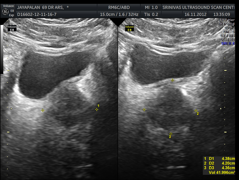
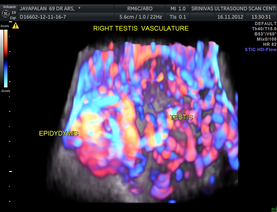
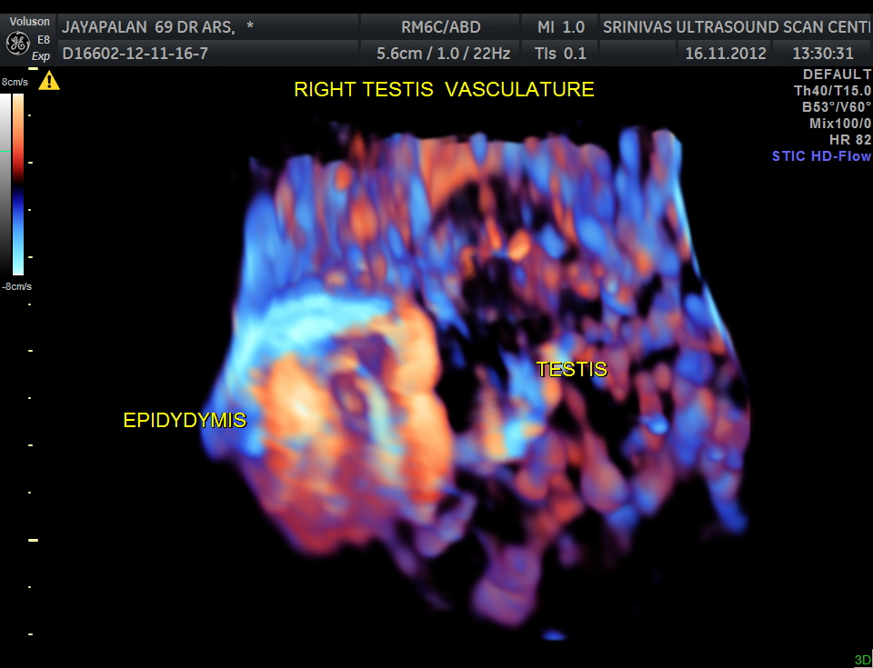
This was a 69 year old gentleman with right scrotal pain and swelling .Scan was requested to differentiate between epidydymo orchitis , testicular torsion or any mass lesion.
Colour doppler gives the answer instantly , Increased vascularity is epidydymo orchitis ; no vascularity with hypo echoic texture is torsion ; mass lesion can have localized increase of vascularity.
The following images were obtained . Power doppler and 3D reconstructed power doppler images show the increased vascularity very well.


good one
LikeLike