
This 29-year-old lady was being investigated for heavy intermenstrual bleeding during her last cycle. She has 2 children. The last child birth was 4 years ago. She gave history of one medical termination of pregnancy between the two children.
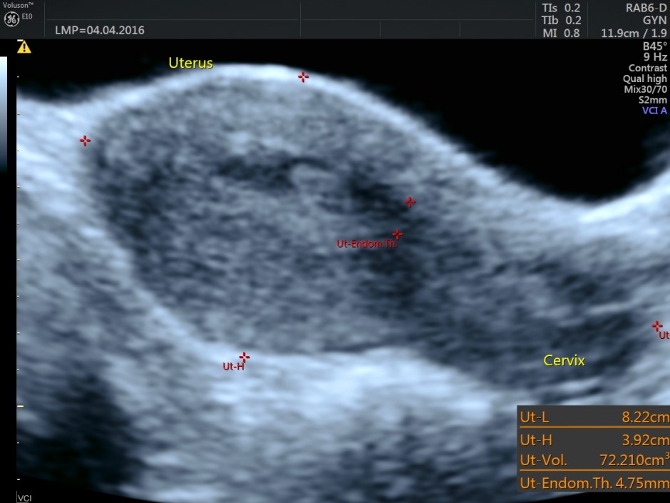
The uterus in 2 D is shown below.
Trans abdominal view
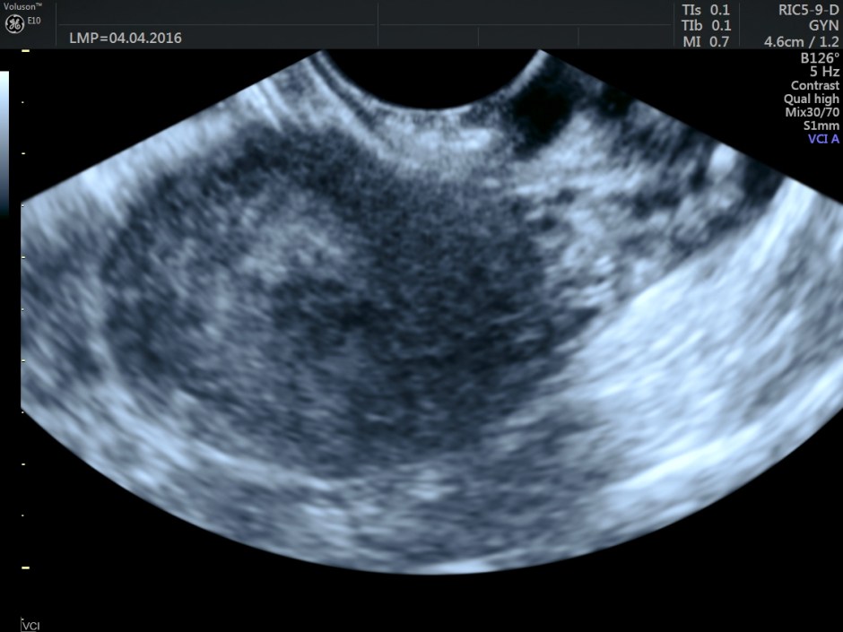
Transvaginal view
Volume contrast imaging pictures are given below:
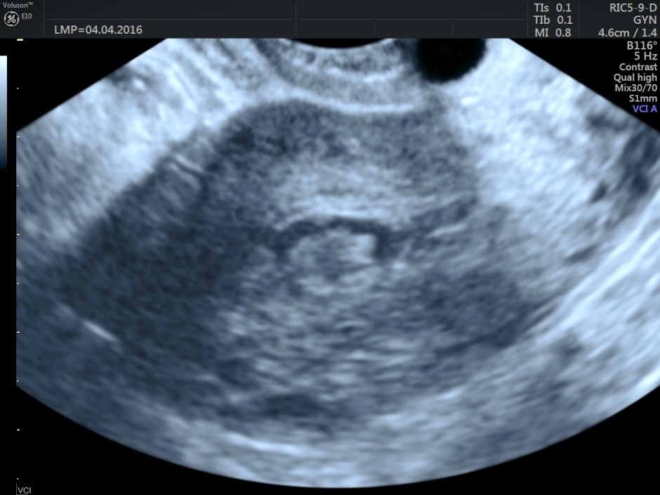
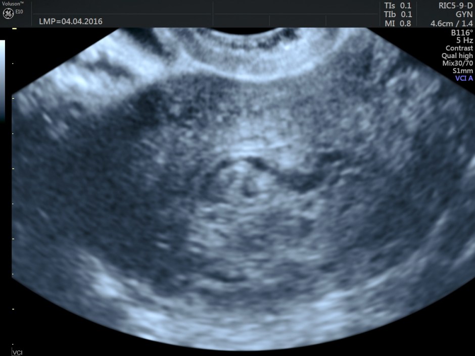
Endometrium shows ? polypoid appearance; Irregular hypoechoic myometrial texture with some serpiginous appearance is seen.
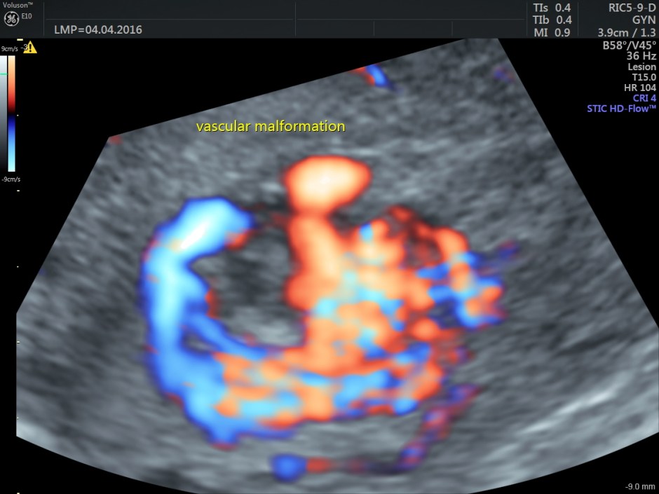
Colour and Power Doppler images are given below.
Trans abdominal :
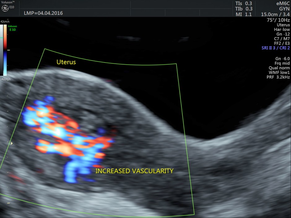
Increased vascularity is seen in the posterior aspect
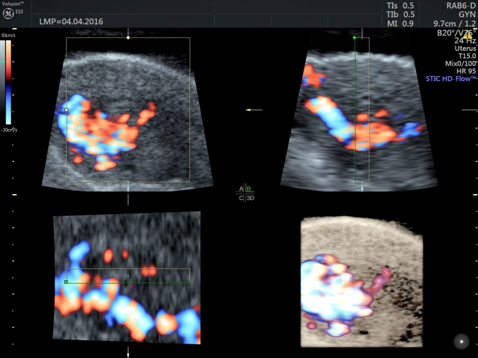
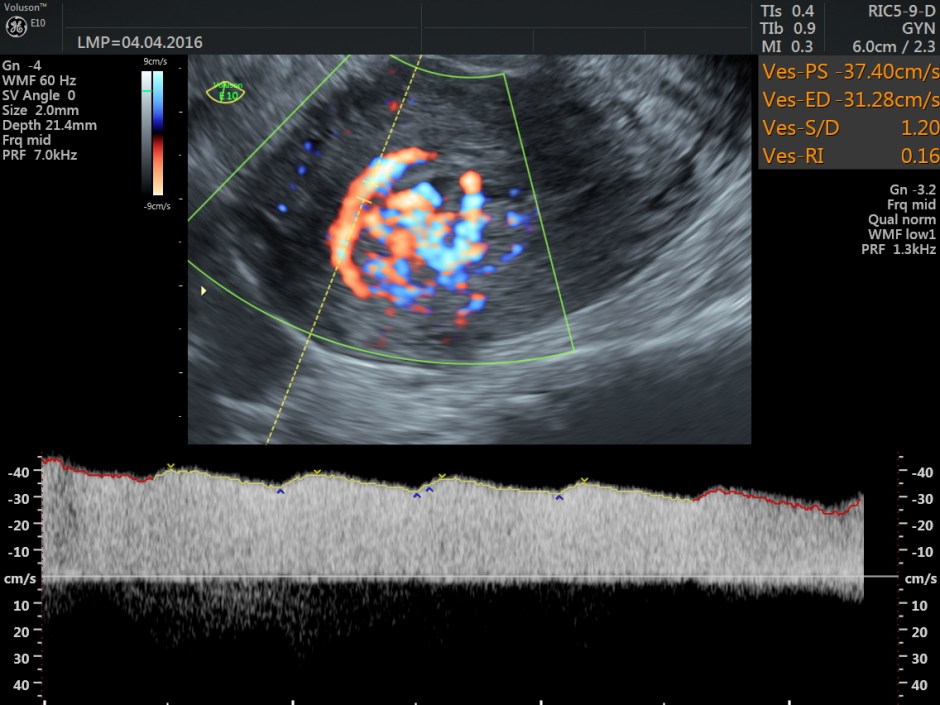
Power Doppler reconstruction images of trans-abdominal scan are given below
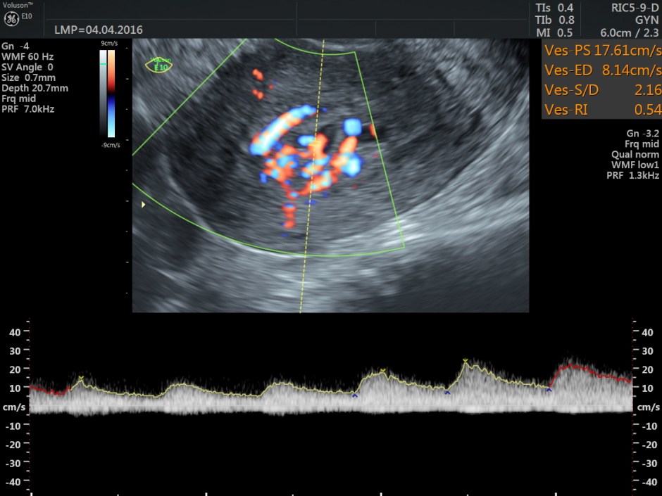
Spectral Doppler images show arterial flow with low resistance at different points .
The following are the different sections of the Power Doppler reconstruction images of the trans-vaginal scan.
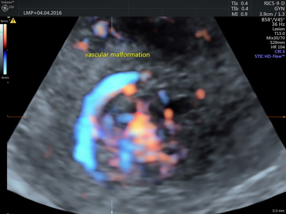
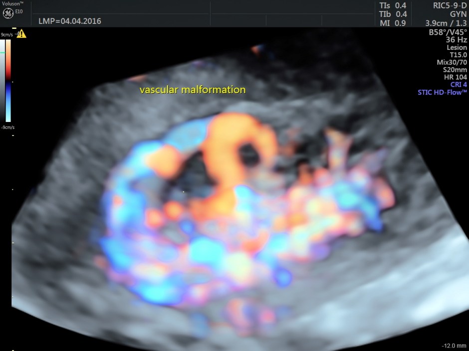
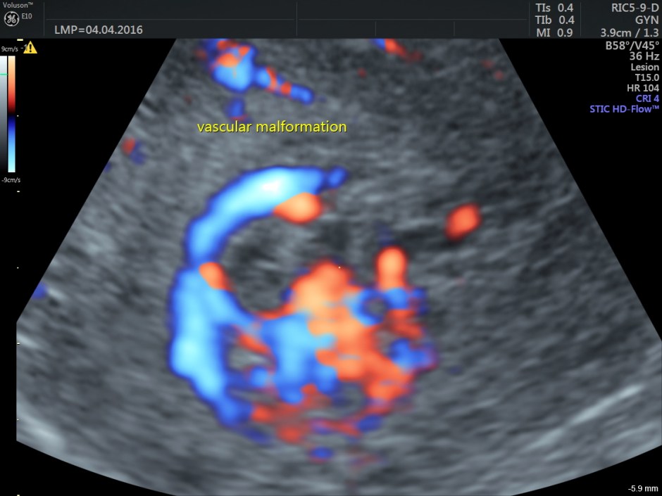
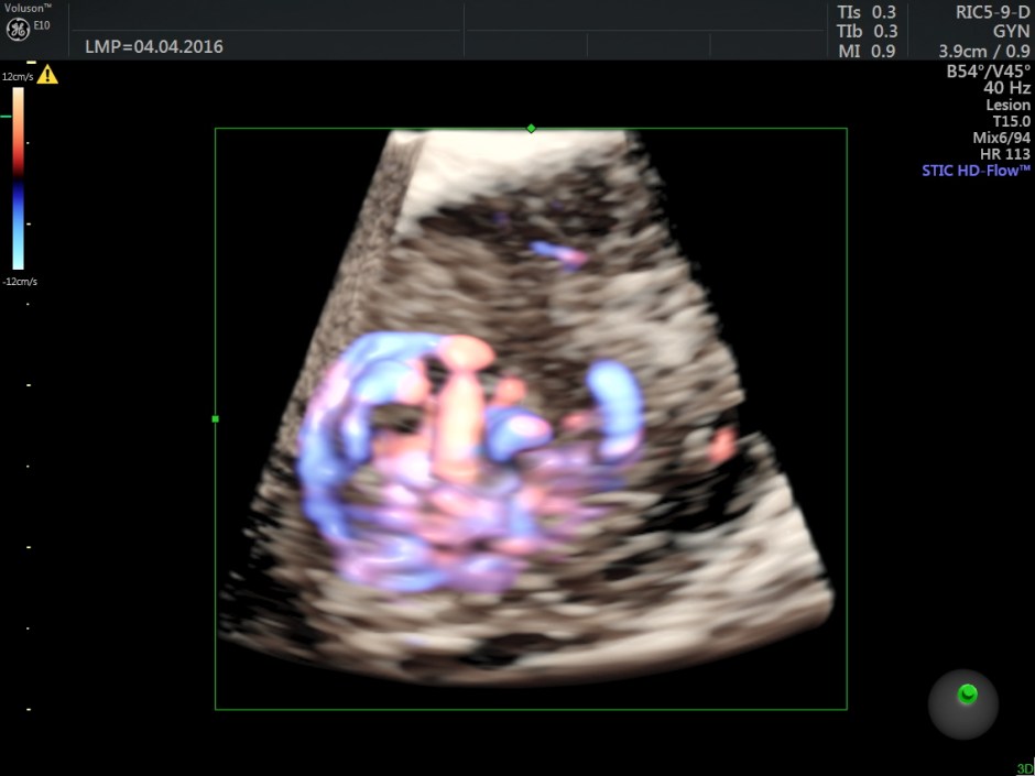
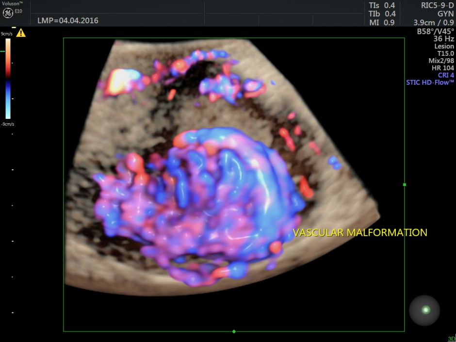
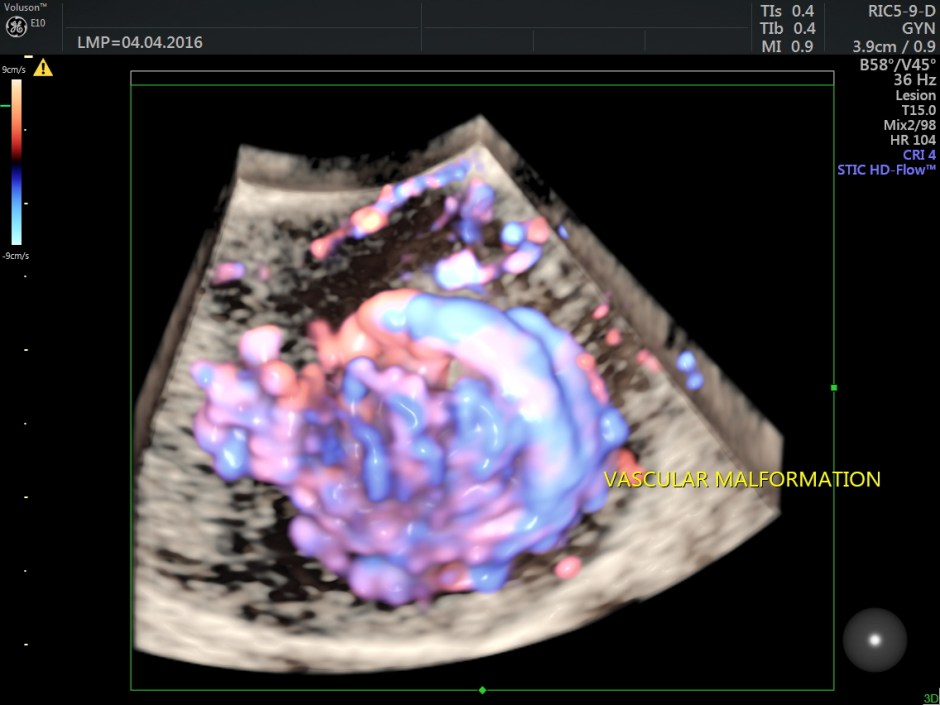
The following are the reconstructed 3D Power Doppler images.
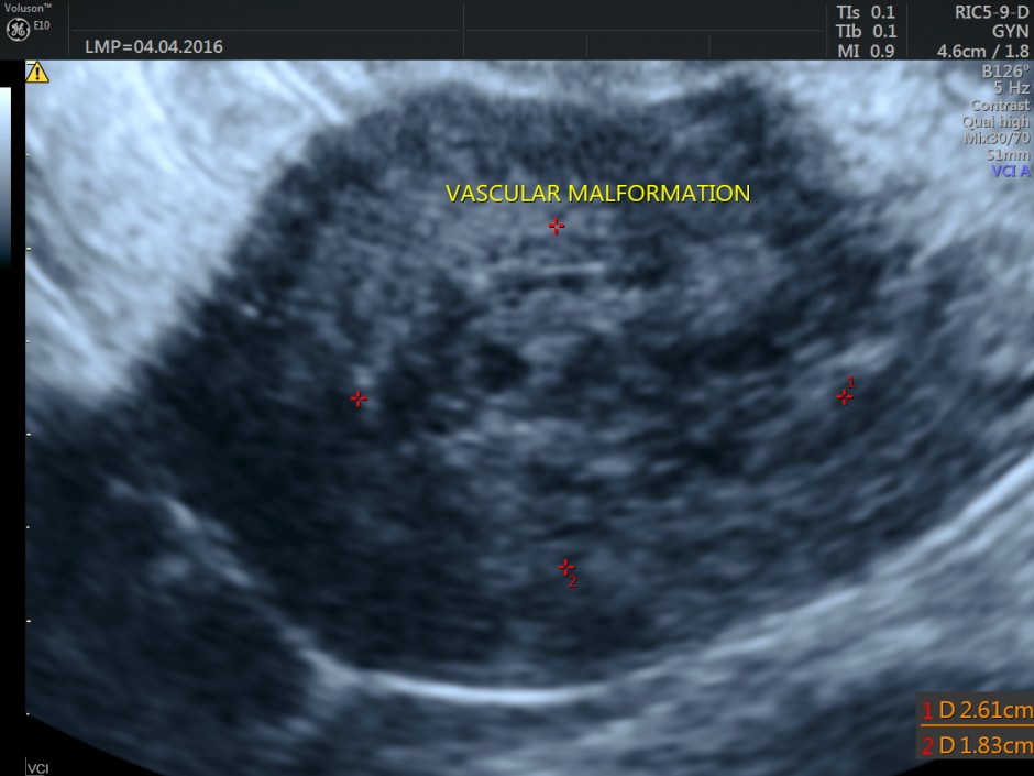
For comparison a 2 d image is given below.
The ultrasound diagnosis was a vascular malformation in the uterus .She was referred to an interventional radiologist,who did a CECT the next day.
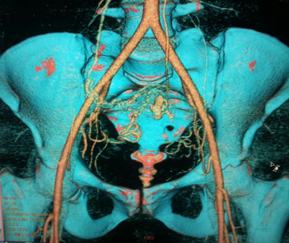
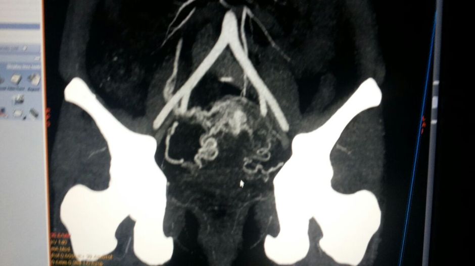
The Contrast Enhanced CT scan pictures are given below.
This was reported as Arterio Venous Malformation, with arterial supply from both the uterine arteries and the venous drainage into the right ovarian vein.
She has been advised embolization treatment of both uterine arteries.
http://radiopaedia.org/articles/uterine-arteriovenous-malformation
http://www.jultrasoundmed.org/content/25/11/1387.long
The following is excerpts from the above:
Uterine arteriovenous malformations (UAVM) result from formation of multiple arteriovenous fistulous communications within the uterus without an intervening capillary network.
Presentation can vary. UAVMs can cause life-threatening massive bleeding in young women. Bleeding is the major presenting symptom in AVMs. As these malformations are less common after menopause, post-menopausal bleeding is rarely seen.
Acquired UAVMs disease are associated with conditions such as :
- multiple pregnancies
- miscarriage
- previous surgery
- dilation and curettage
- termination of pregnancy
- caesarean section
Gray-scale sonographic appearances can be non-specific and can have a range of manifestations including areas of subtle myometrial inhomogeneity, tubular spaces within the myometrium, a intramural uterine, endometrial or cervical mass like region or sometimes as prominent parametrial vessels . The extent of mass is effect is however minimal.
Colour Doppler
Typically shows serpiginous/tubular anechoic structures within the myometrium with a low resistance (RI ~0.2-0.5), high velocity flow pattern on colour Doppler interrogation.
Treatment
Dilatation and Curettage should be avoided as it may lead to catastrophic bleeding.
Transcatheter arterial embolisation is an excellent treatment option in selected cases.
Earlier hysterectomy was advocated.
Endovaginal sonography is the imaging modality of choice in patients with abnormal uterine bleeding.
Routine use of color and spectral Doppler sonography allows one to confidently make the correct diagnosis.
Transcatheter arterial embolization is an excellent treatment option.
Endovaginal sonography should be used to monitor postembolization outcomes.

Very interesting case nicely resolved. Thank you very much for sharing
LikeLike
Thank you
LikeLike
Another interesting case! Indeed a perfect demonstration for the use of colour techniques. Thank you Kriznan.
LikeLike
Thank you
LikeLike
Quite interesting and great demonstration of malformation as they are not as common and not as extensive. Thanks for posting.
LikeLike
Thanks
LikeLike