This week we have a few random 3d reconstructed images .
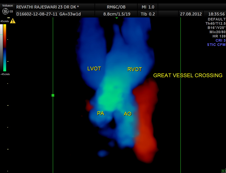
first is a great vessel crossing ; ideally the fetus should be on its back facing the examiner .and should be still for the duration of the 3 D sweep.This view , if normal essentially rules out transposition of great vessels.
the next is a 3 d reconstruction of fetal pulmonary vasculature.
viewing atleast one o r two pulmonary veins draining into the left atrium effectively rules out total anomalous pulmonary venous drainage
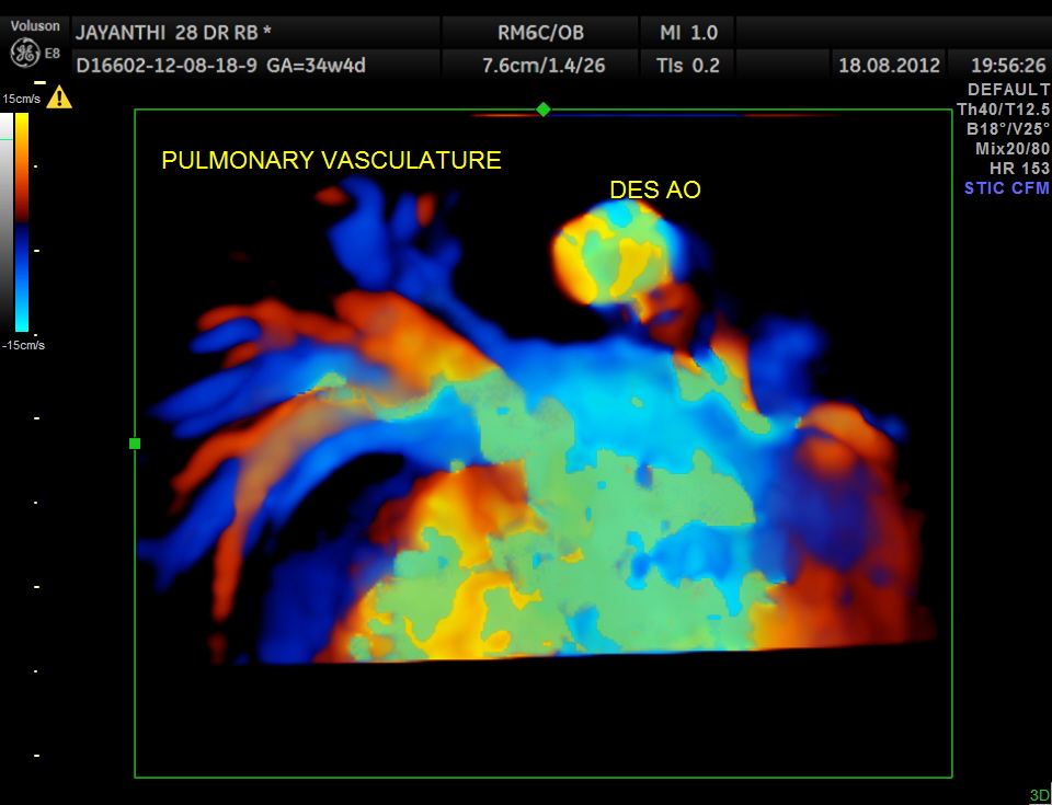
all 4 pulm veins are seen in this reconstruction
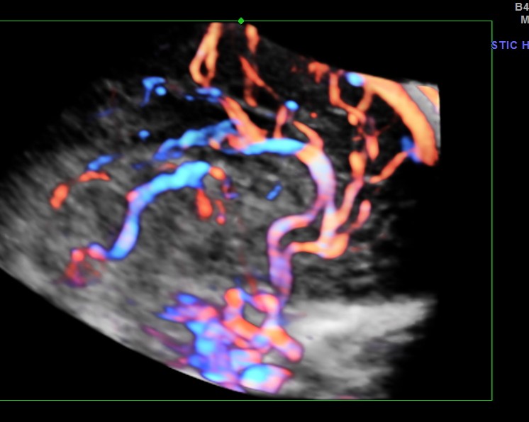
the next is a reconstruction of pericallosal artery
.viewing the peri callosal artery in full implies that the corpus callosum is formed normally.

the next is a reconstruction image of aortic arch

