This was a 27 year old lady , Gravida 2, Para 1 , Live 1 , Miscarriage 0.
The scan was done around the 35 weeks.The following images were seen .
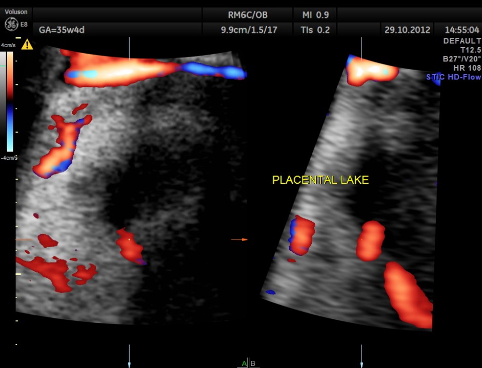
apart from this the placenta showed a few lakes and a localised mass , probably chorio angioma
 No other fetal anomaly was made out
the important views of the heart are shown below
fetal face
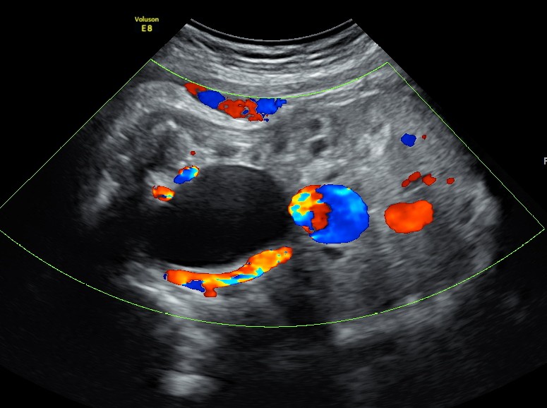
 The differential diagnosis : urachal or ovarian cyst ; easily differentiated with the Doppler examination.
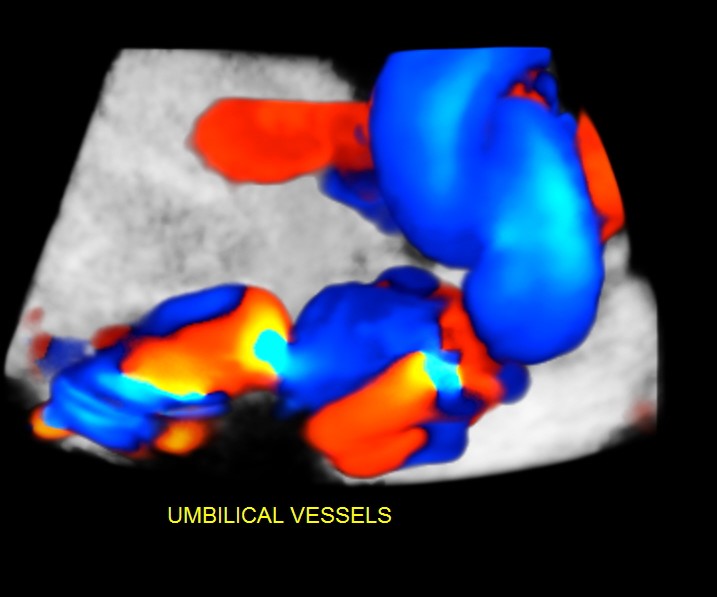
An umbilical varix is a developmental rather an embryologic abnormality and most cases have a normal ultrasound at 16 to 19 weeks gestation.
Unlike persistent right umbilical vein, umbilical vein varices have not been associated with other congenital malformations.
The significance of an antenatally detected umbilical varix remains controversial.
This finding has been associated with an unexplained high mortality rate in utero: thrombosis of the dilation leading to fetal death and other complications including hydrops fetalis. It had also been linked with chromosomal abnormalities.
Karyotyping is not warranted if not any associated malformation is detected.
When a fetal intra-abdominal umbilical vein varix is isolated, a good fetal outcome is expected.


Nice you left a comment at the bottom ,so I could understand ,great! Narayanan
LikeLike
Thanks
LikeLike