This was a 36 year old primi , non consanguinous ; she gave h/o taking sodium valproate for seizure disorder for the last 10 years.
The scan was done around 25 weeks of gestation :

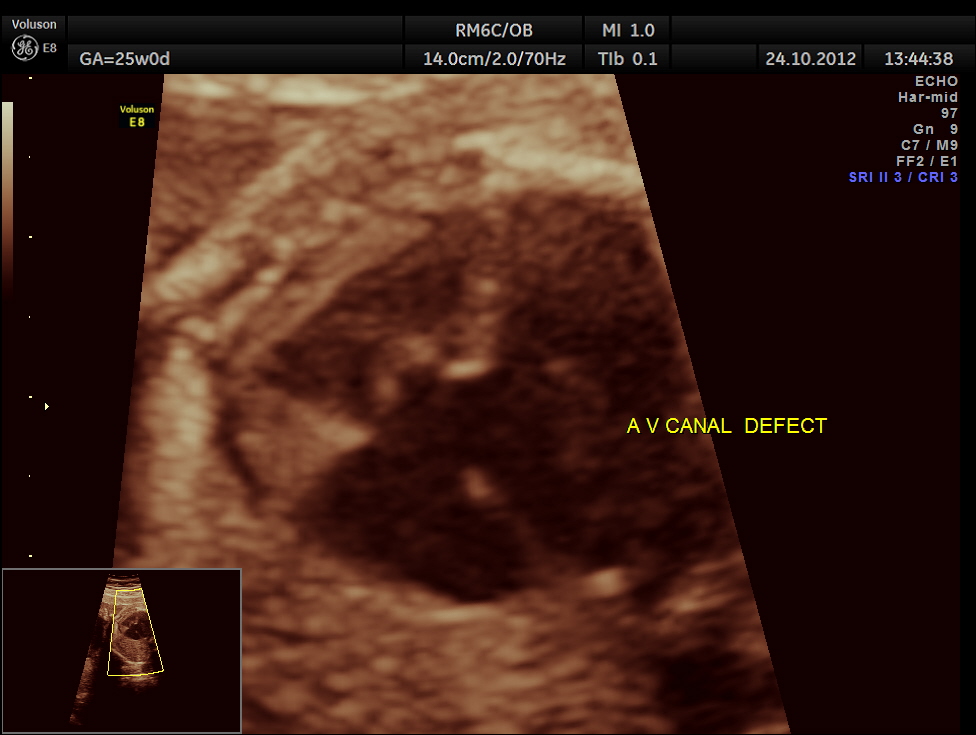
prominent endocardial cushion defect or a v canal defect seen

colour flow clearly demonstrating the atrio ventricular septal defect

Spectral doppler shows high velocity flow across the defect


normal situs seen

Left Ventricular Outflow Tract view ( LVOT )

3 Vessel view
the following are spectral doppler flow across the 4 valves

mitral valve

aortic valve

pulmonary valve

tricuspid valve
the last is a reconstructed image of the aortic arch

prognosis depends on type
http://ats.ctsnetjournals.org/cgi/reprint/2/3/399.pdf
Type I. Persistent ostium primum without significant deformity of atrioventricular valves
Type 11. Persistent ostium primum with cleft of the atrioventricular valves
Type 111. Persistent ostium primum with cleft atrioventricular valves and subjacent ventricular septal defect
Type IV. Same as 111, but with pulmonary hypertension at or near systemic levels
Type 0. Those types not included above
Type I and Type I1 are self-explanatory and include the most com-
mon surgically encountered variants of endocardial cushion defects.
Types 111 and IV divide the complete form- of persistent atrioventric-
ular canal on a physiological basis. This division is clinically essential,
since the very high surgical mortality of Type IV lesions sets them apart

Your blogs r nice and usefull!
LikeLike
thanks for the comment. 🙂
LikeLike