This was a 55 year old gentleman, with complaints of dyspepsia , belching and vague abdominal pain of 4 months duration. There was no history of any fever. He was a non smoker and gave history of occasional alcohol consumption. He was working in the middle east and had come to India for evaluation and treatment.
He was seen by a surgeon, who asked for an ultrasound . This was reported as abscess in the right lobe of the liver. He was referred to a medical gastro-enterologist , who asked for a CT scan and it was reported as hypo dense lesion in segment 8 of the liver – probable abscess. As it had a volume less than 40 cc , medical management with antibiotics was given and he was not getting better. He came to our out patient department and he was subjected to a repeat ultrasound.
The following pictures were obtained.
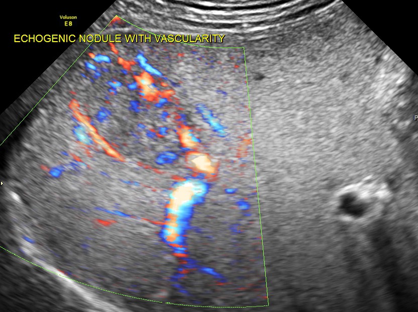
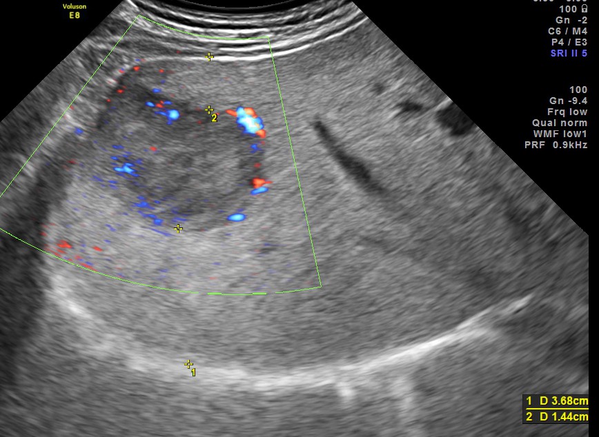
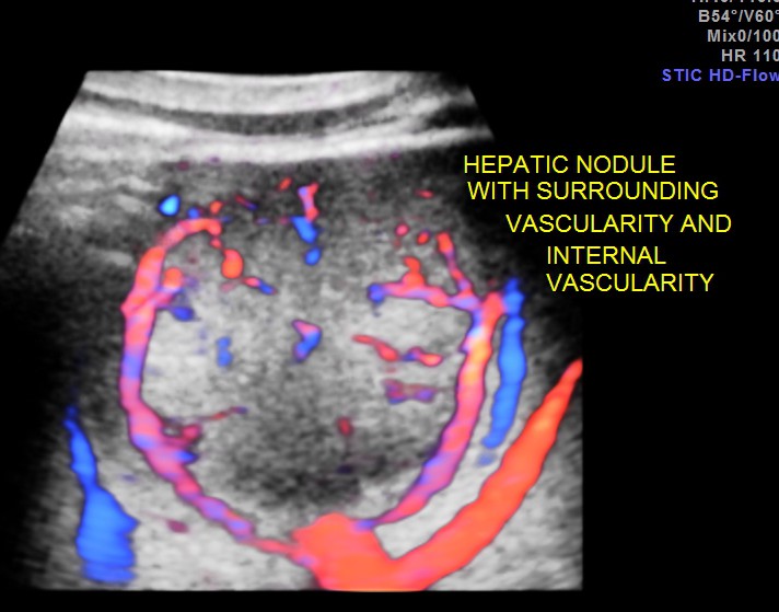
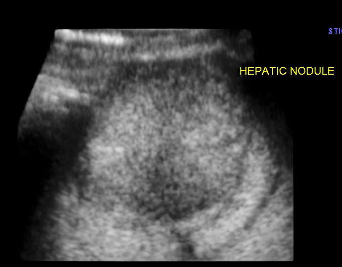
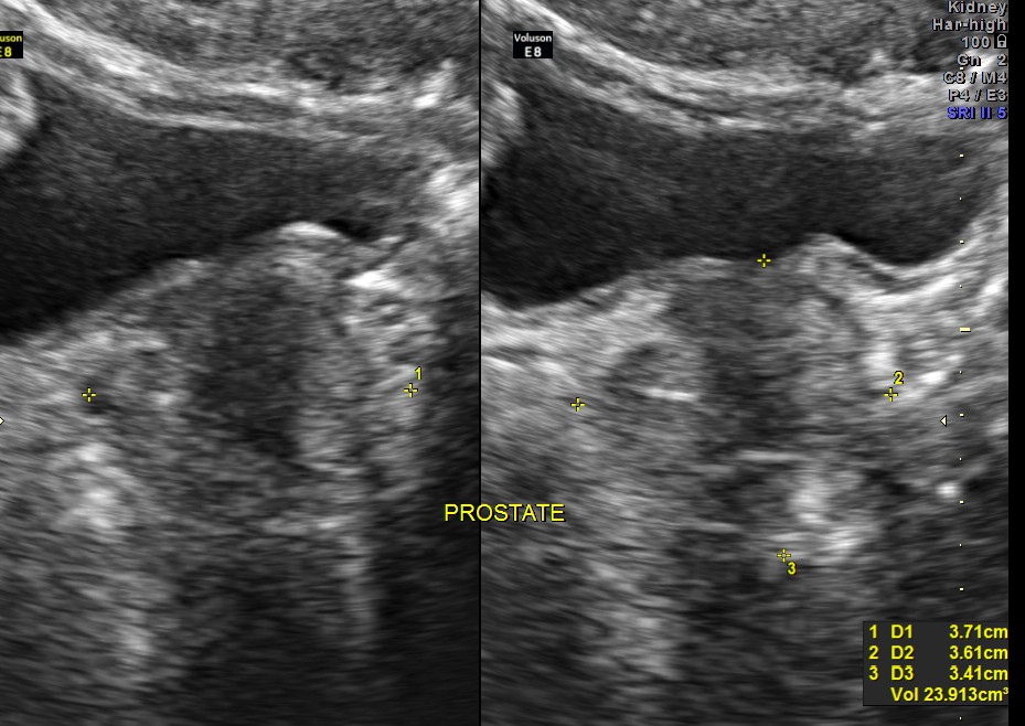
An echogenic nodule with vascularity is seen.
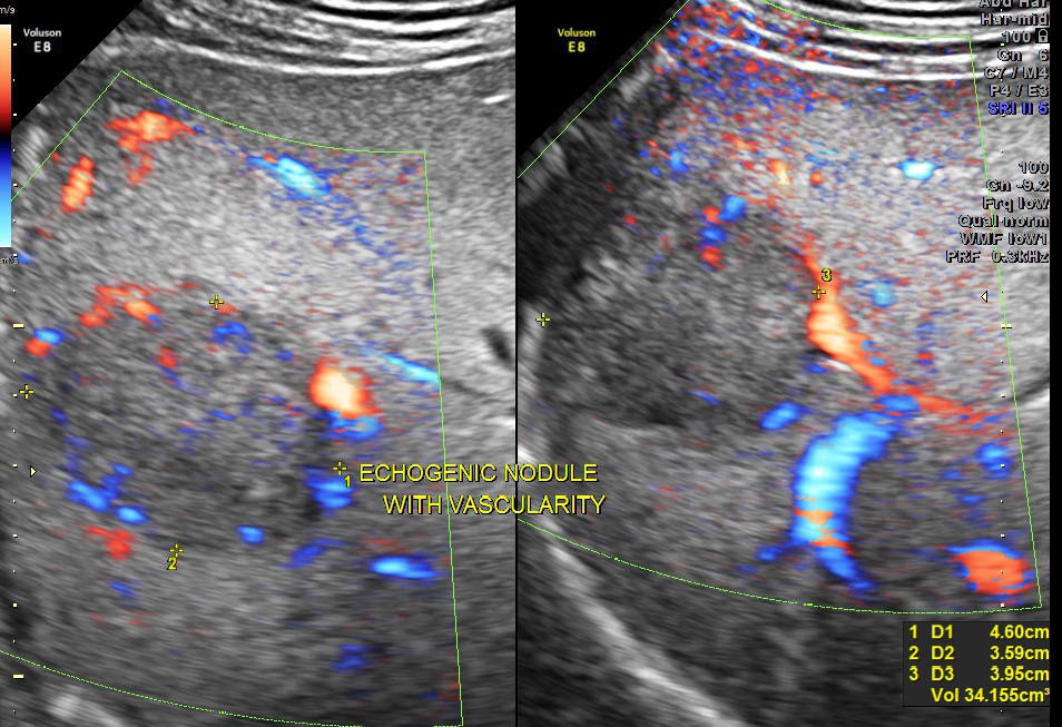
3 d reconstruction shows the solid nature and vascularity around and within the nodule.

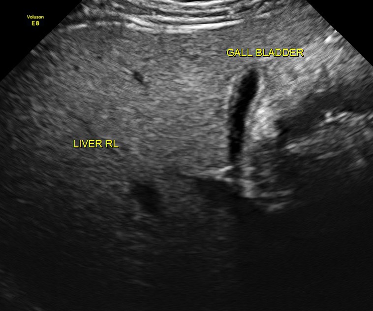
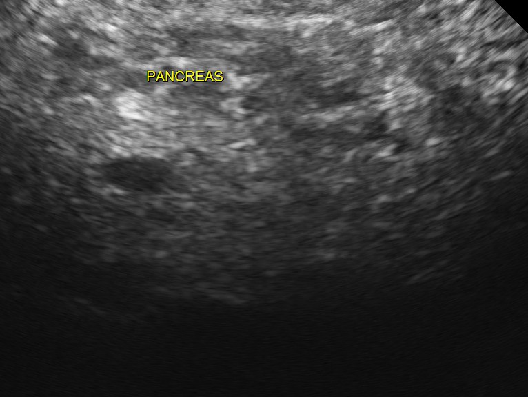
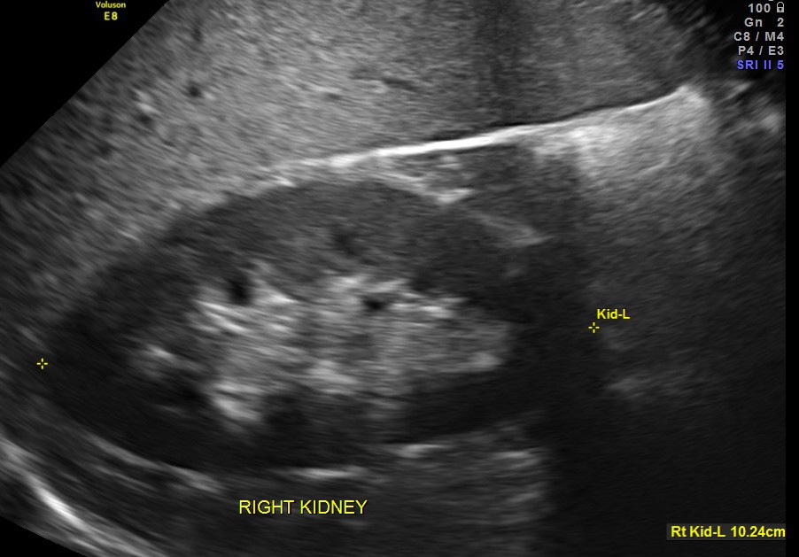
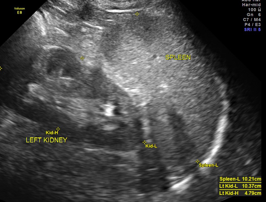
The scan was otherwise normal.
The appearance of this nodule was suggestive of a metastatic nodule with the increased vascularity around and within.
An upper G.I.Endoscopy was performed the next day and revealed a primary gastric antral carcinoma.
This case is presented here to underline the point that colour and power doppler can help us in deciding correctly about a gray scale finding which could signify multiple diagnoses.

GREAT
LikeLike
interesting
LikeLike
We reported two cases of Liver abscesses burst through anterior abdominal wall and right dome of diaphragm both mimicked neoplasm in the ASEAN J Radiology in 2003.
LikeLike