
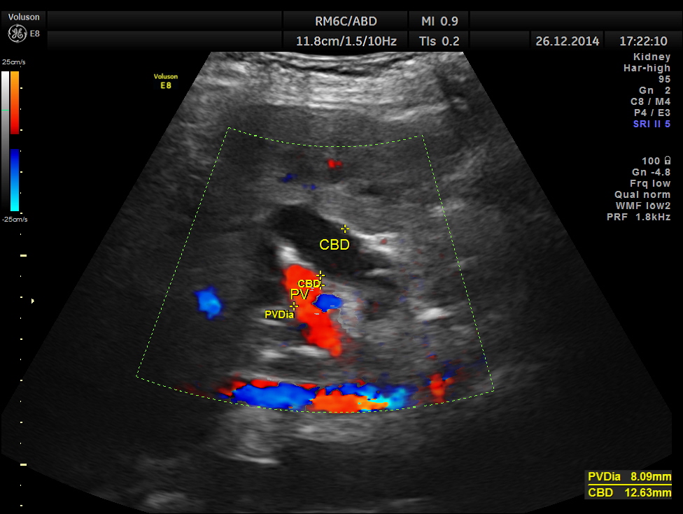
This was a 82 year old lady being evaluated for abdominal pain . Her serum bilirubin was very high and she was referred for an ultrasound .
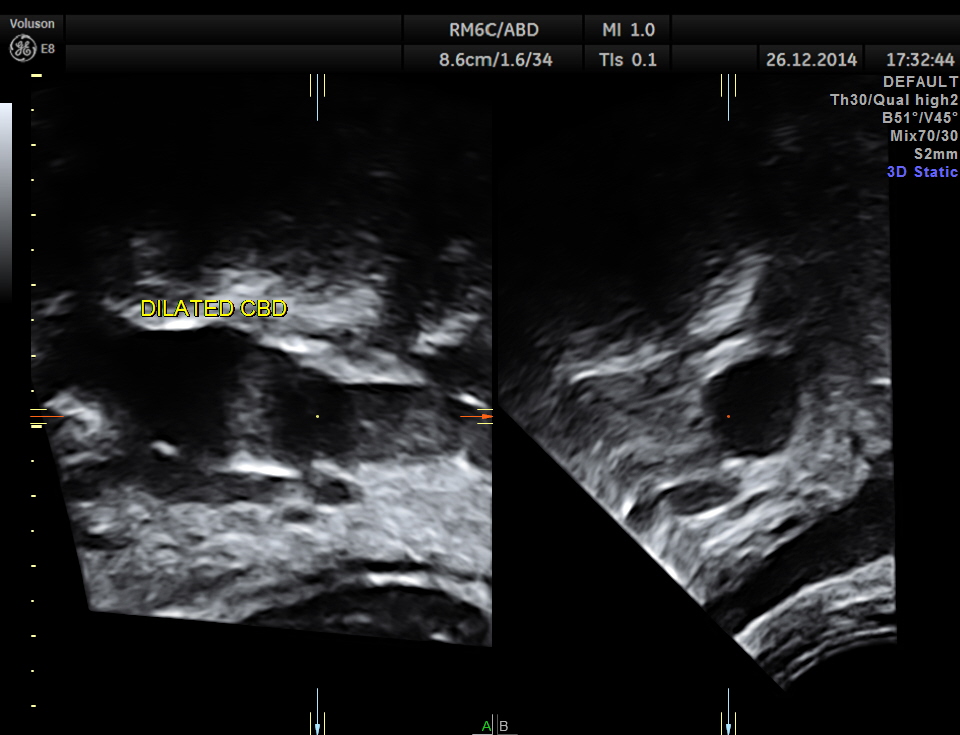
grossly dilated common bile duct.
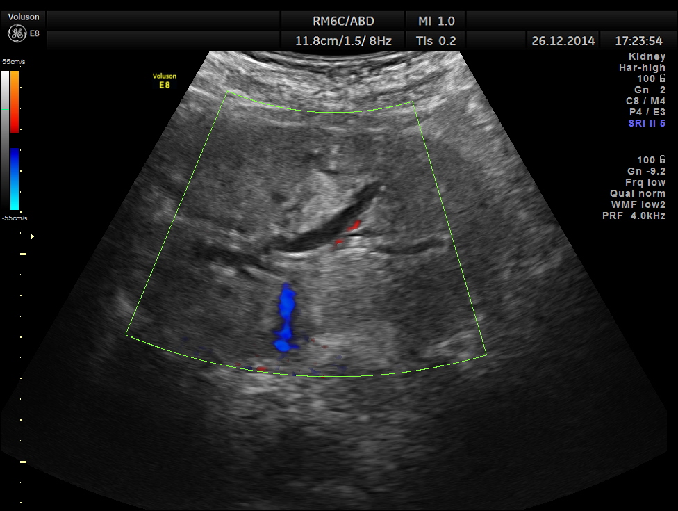
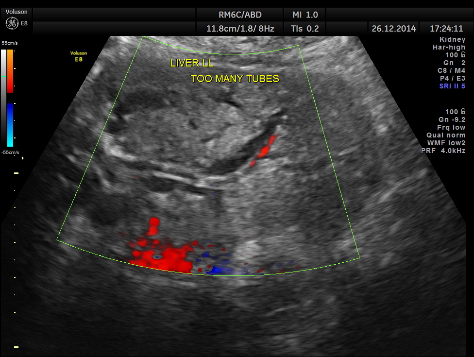
too many tubes seen in the left lobe of the liver.
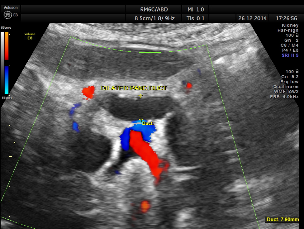
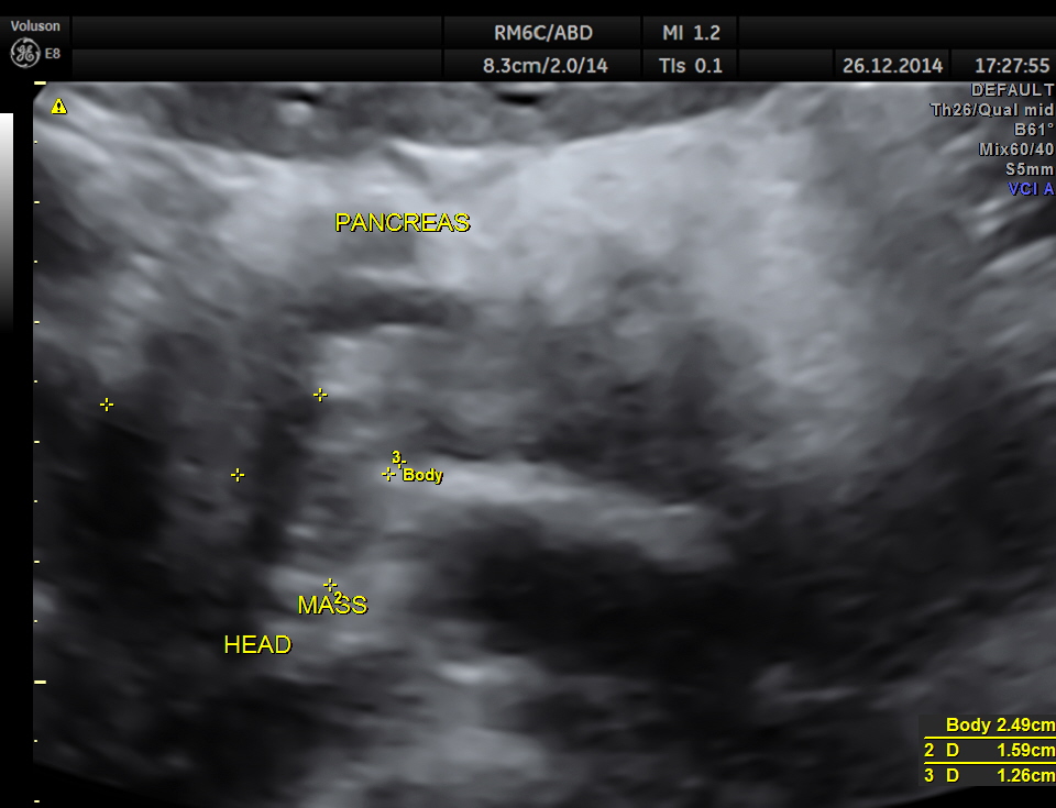
dilated pancreatic duct is seen
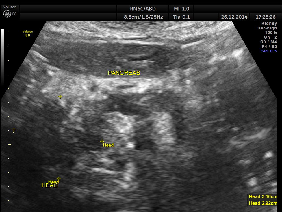
the head region of the pancreas shows an irregular mass lesion
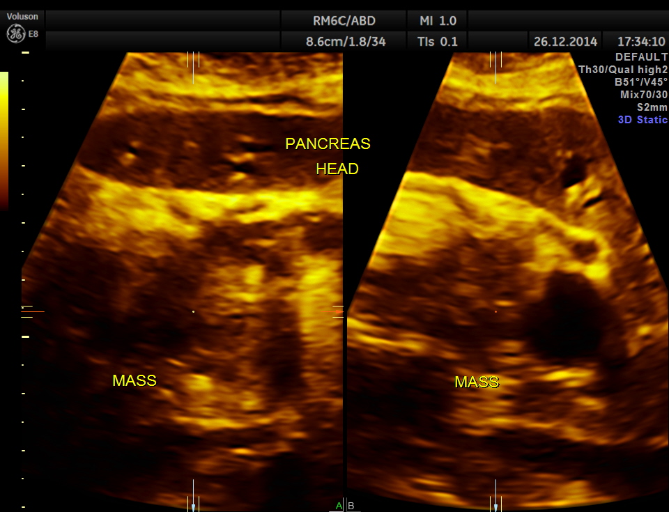
3d reconstruction of the head of the pancreas
3 d reconstruction of extra hepatic CBD
volume contrast image – A plane image of the pancreas
free fluid seen in the abdomen and pelvis , suggestive of possible peritoneal spread of Ca.
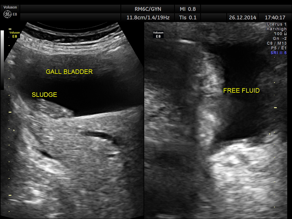
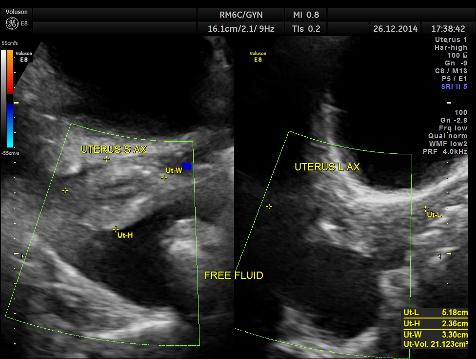
This was a case of Carcinoma of the head of the pancreas , causing biliary obstruction and also pancreatic ductal obstruction , with possible peritoneal deposits.

Thank you Kriznan
LikeLike