
This was a 48 year old gentleman , who was evaluated for generalised tiredness. His complete blood count showed pancytopenia and clinically he had mild splenomegaly.
Ultrasound examination revealed the following findings :
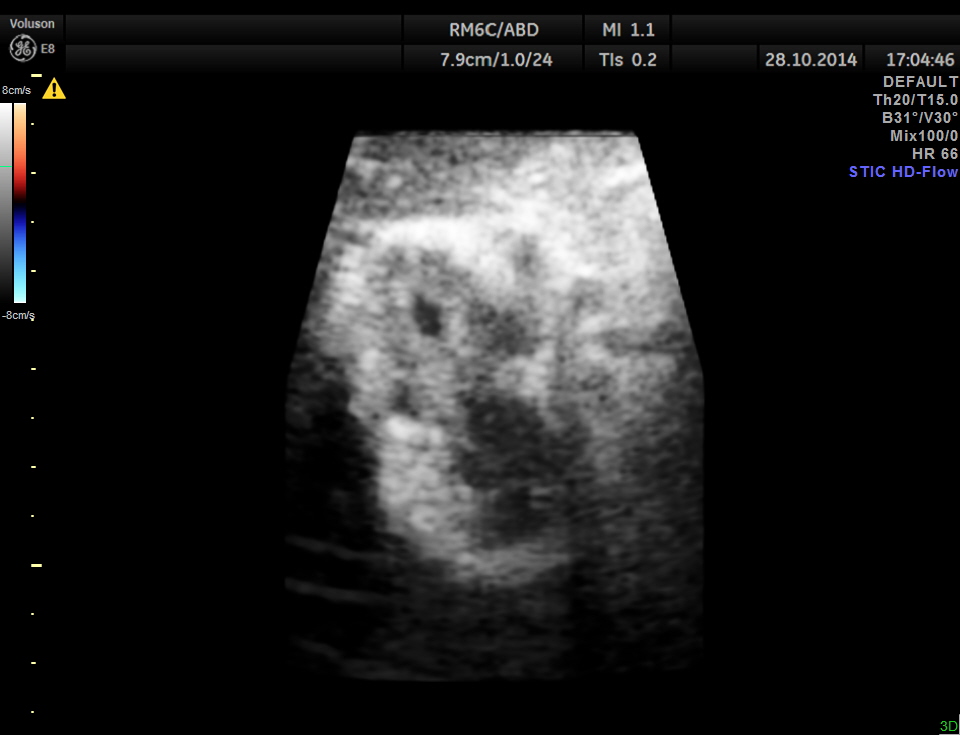
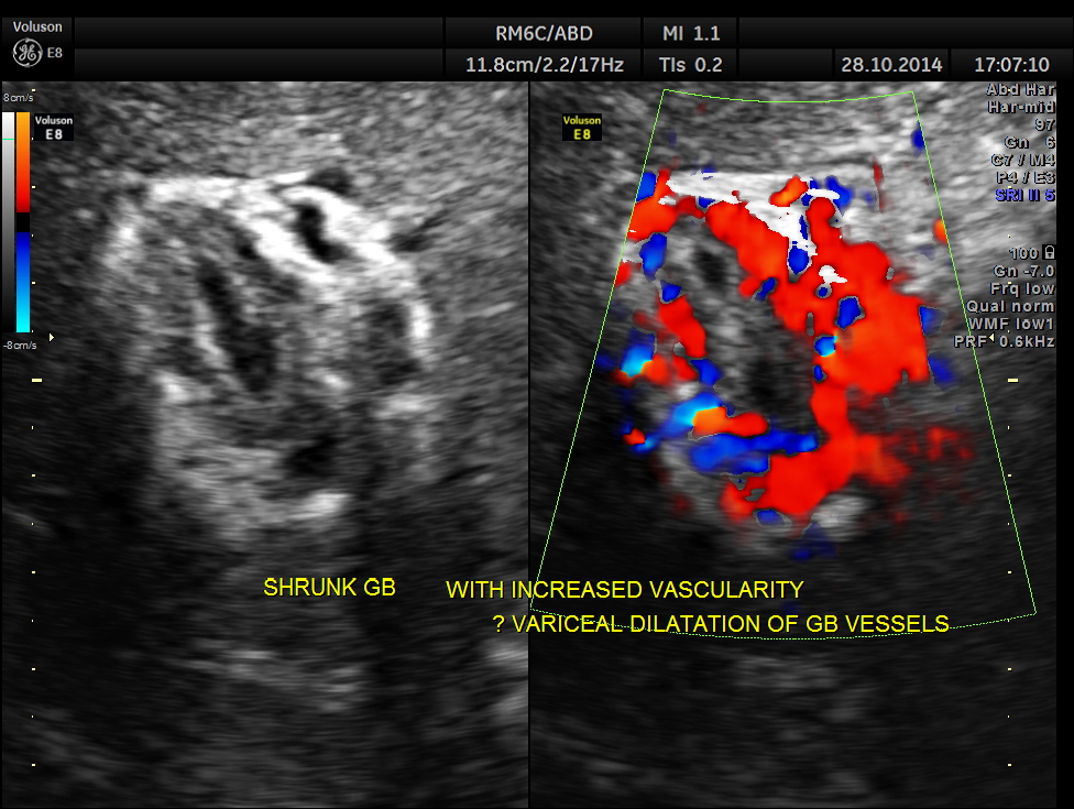
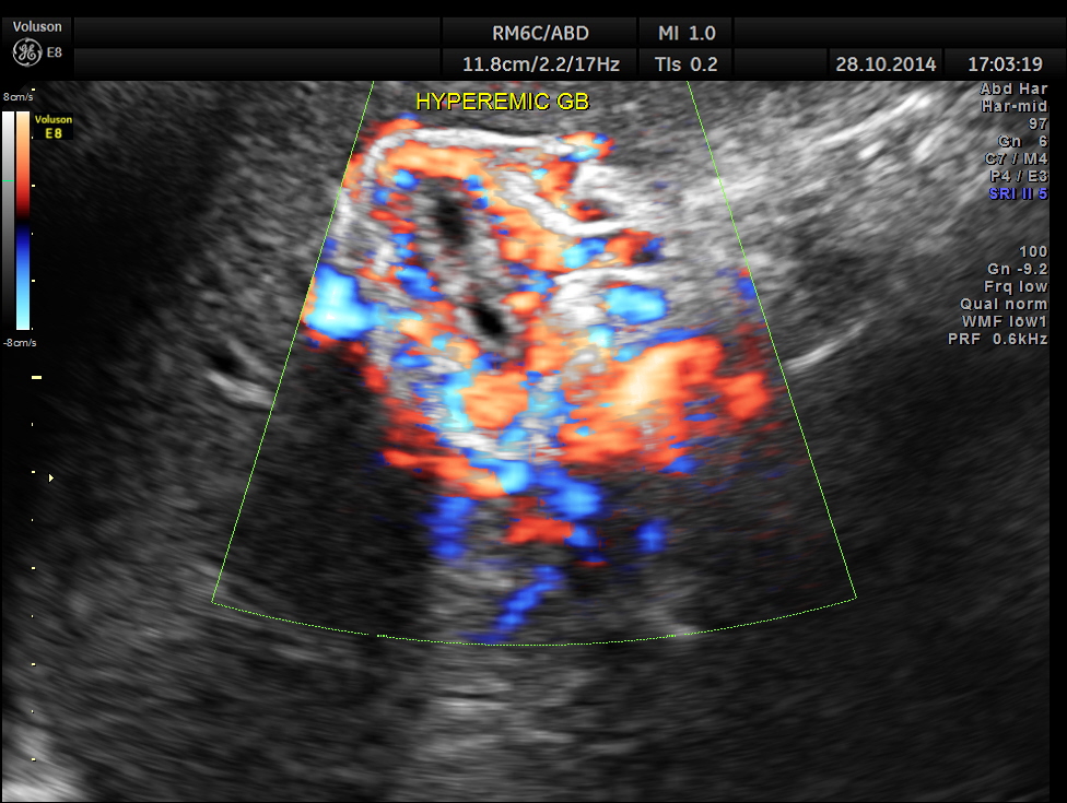
Hypo echoic linear spaces are seen around the gallbladder.
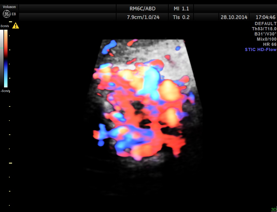
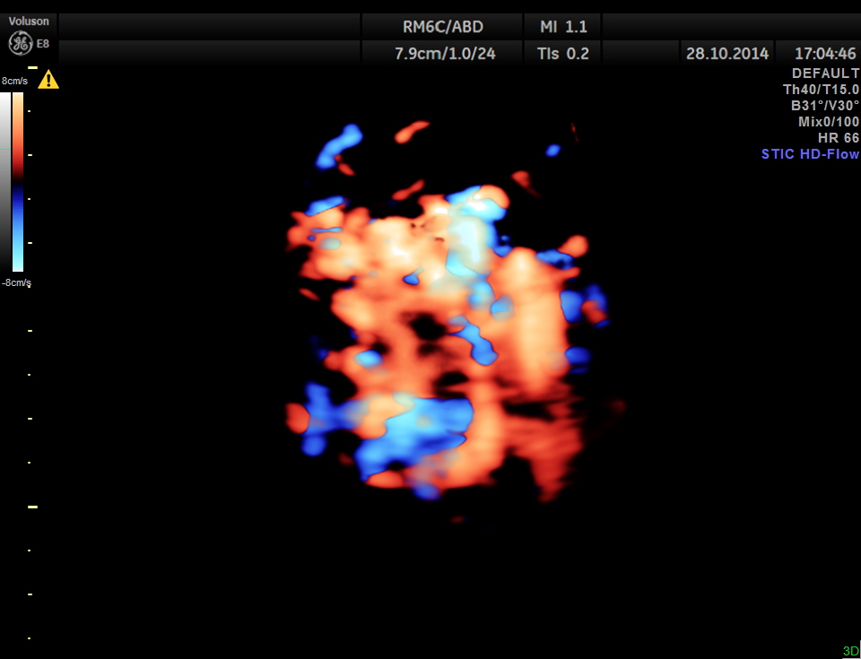
Colour flow imaging of the same
STIC HD flow image.
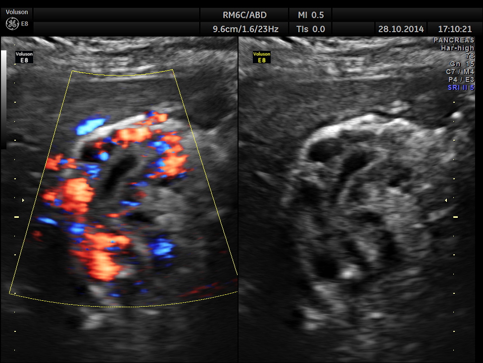
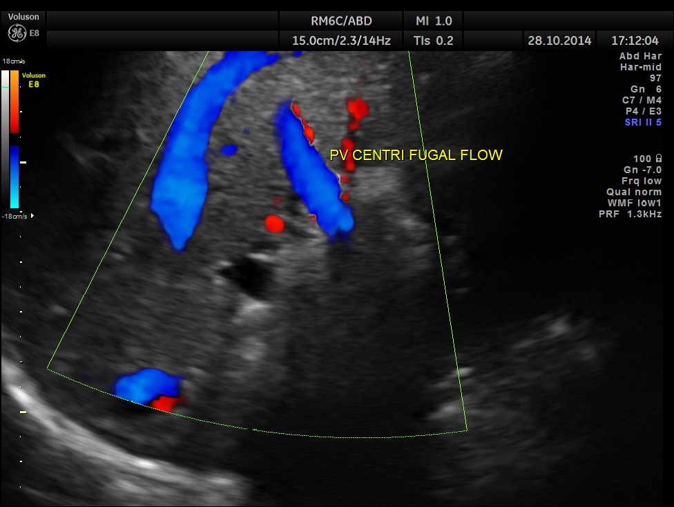
Main portal vein show flow reversal ( blue colour ) – centri-fugal flow suggestive of portal hypertension.
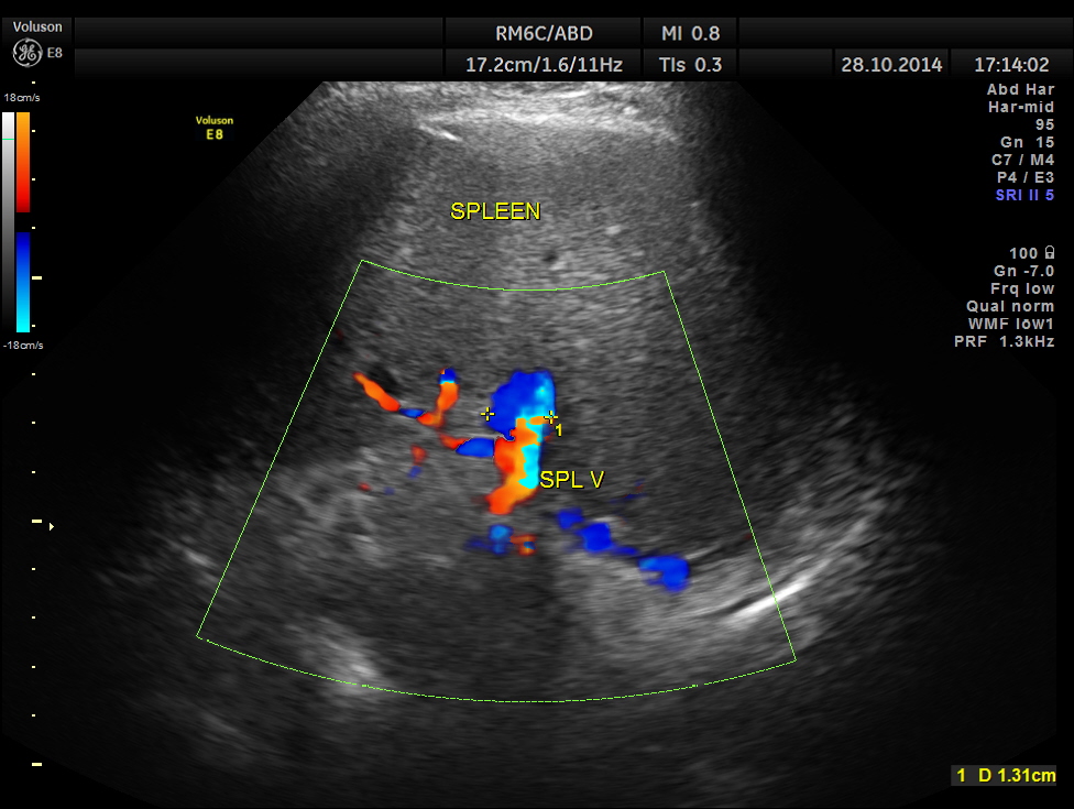
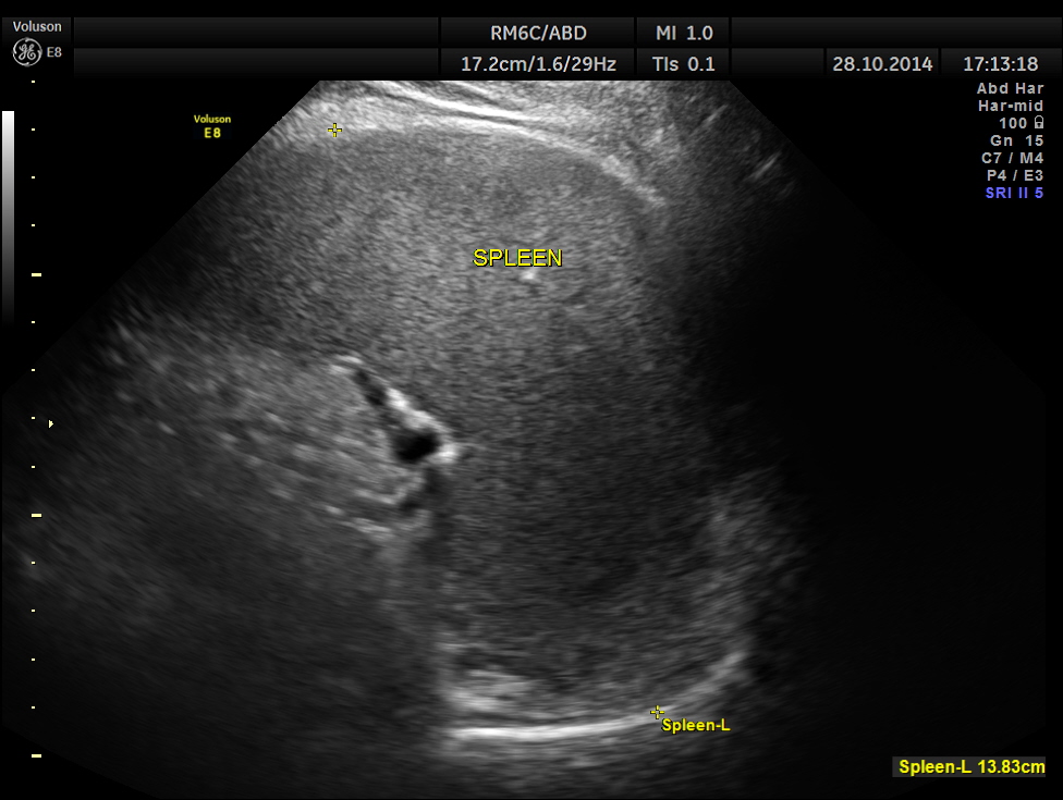
Mild splenomegaly with mildly dilated splenic vein is seen.
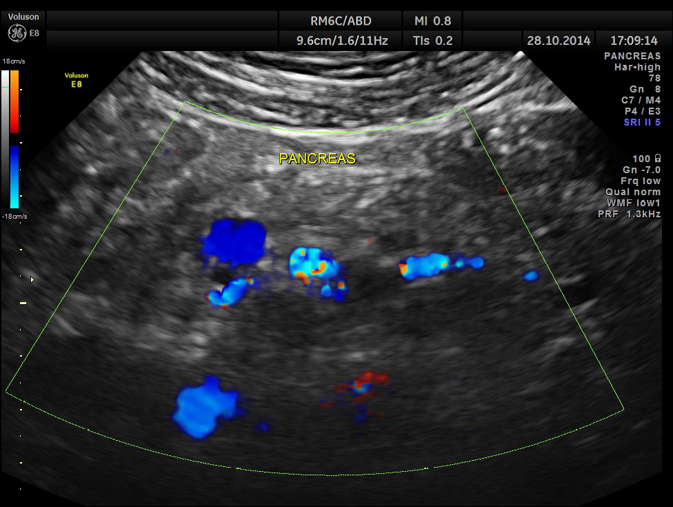
Epigastric varices are also seen.
This patient has cirrhosis of liver with portal hypertension – flow reversal of main portal vein , splenomegaly , mildly dilated splenic vein and varices around the gallbladder.
