This was a 55-year-old gentleman who was being evaluated for painless hematuria.
He had incidental gallstones.
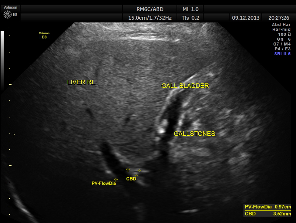
how many gallstones are seen ?
3 d of the same is given below.
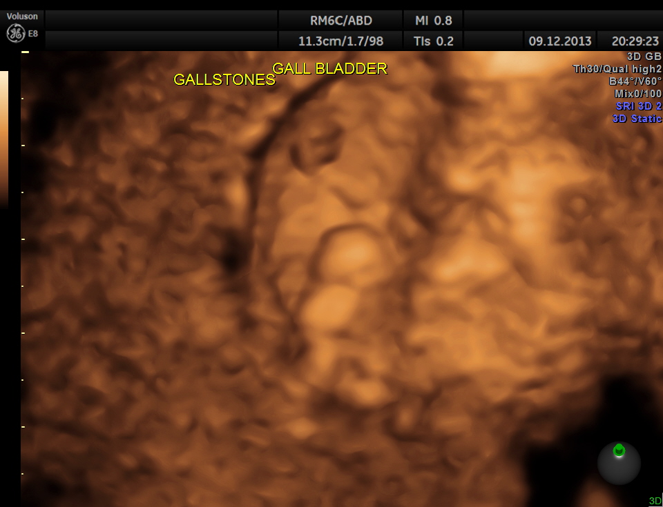
how many are seen ?
high-definition rendering is given below.
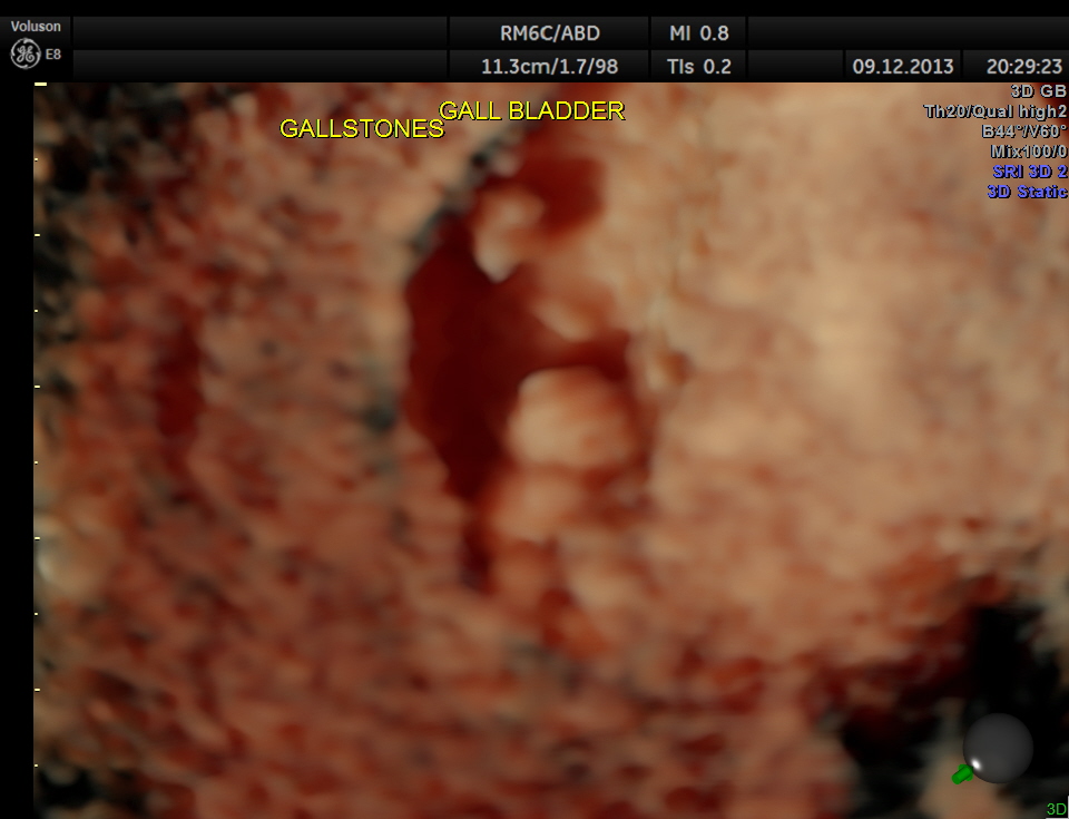
what would be the count now ?
Usually the patient and the surgeon are interested in knowing the number of gallstones . I always have felt that the number would not matter . But is there a difference between 2D , 3D and more advanced high-definition rendering ? Please decide for yourself.
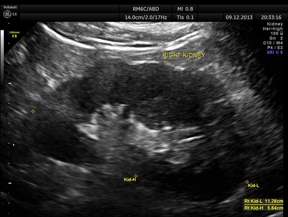
This patient was being evaluated for painless hematuria.
His right kidney appeared normal.
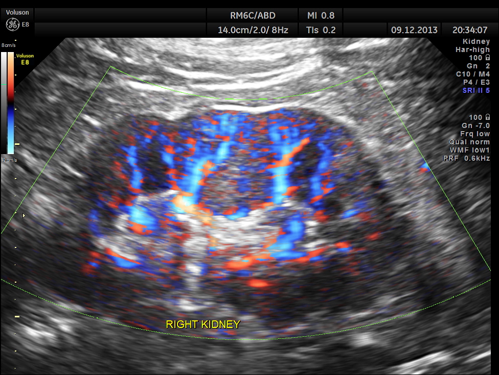
Power Doppler of the right kidney.
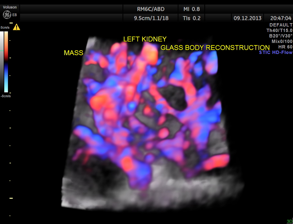
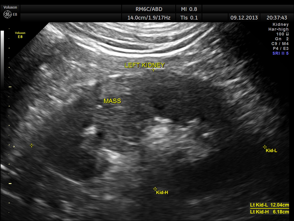
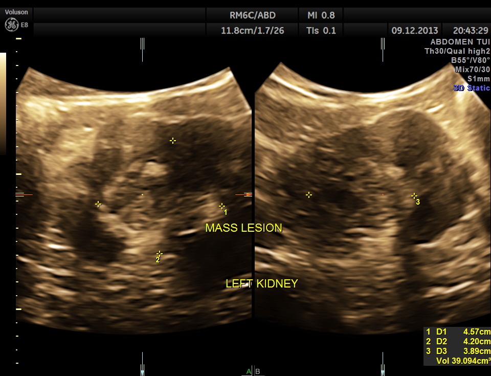
The pictures of the left kidney are given below. An echogenic mass lesion is seen .
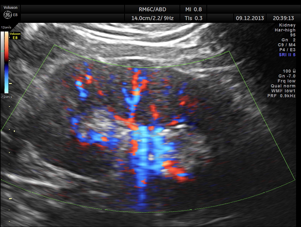
Regular power doppler appears unremarkable.
3D View of the mass.
GLASS BODY RECONSTRUCTION IS GIVEN BELOW.
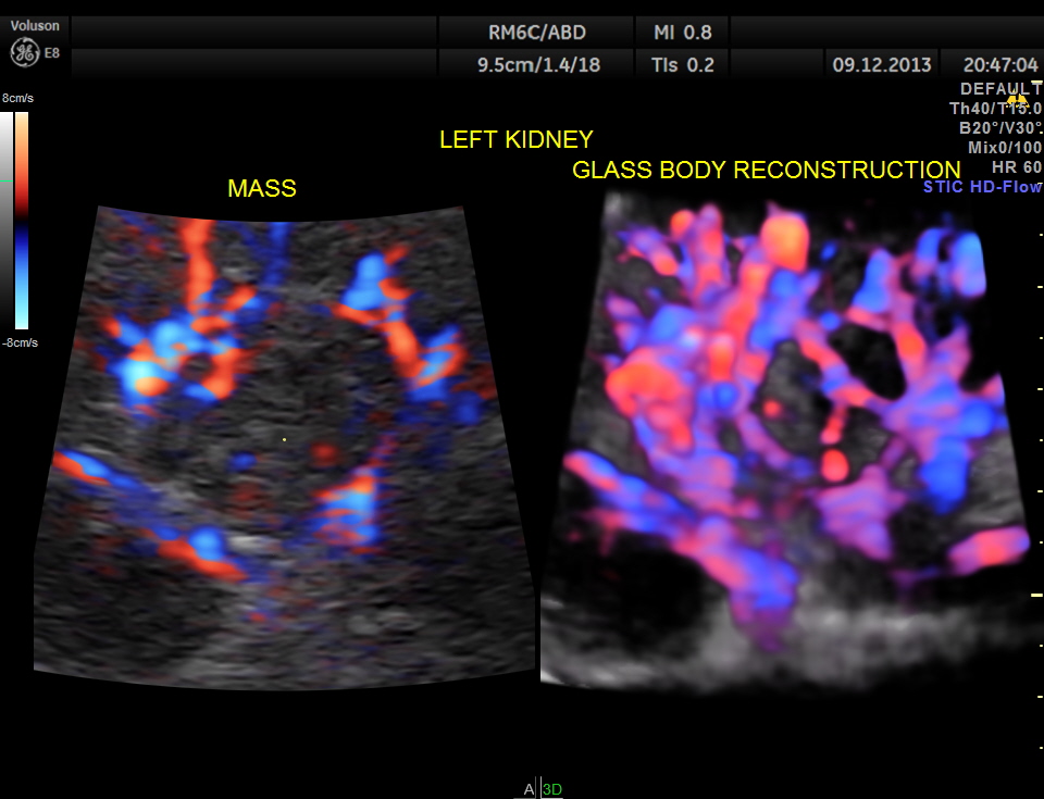
INCREASED VASCULARITY OF THE MASS IS WELL MADE OUT IN THIS GLASS BODY RECONSTRUCTION.

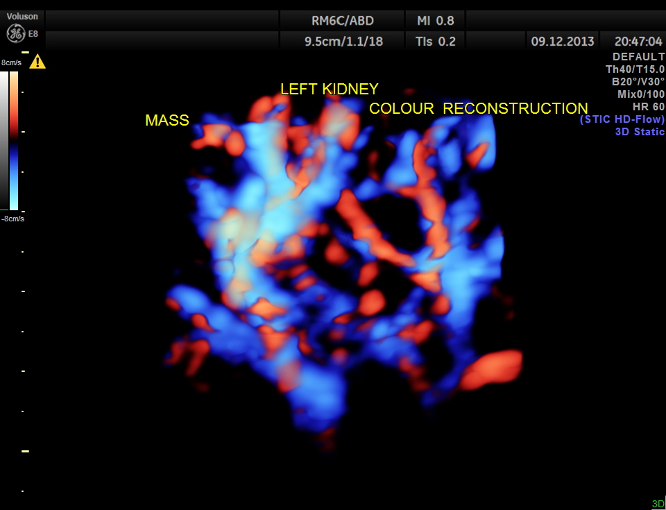
COLOUR FLOW ONLY RECONSTRUCTION.
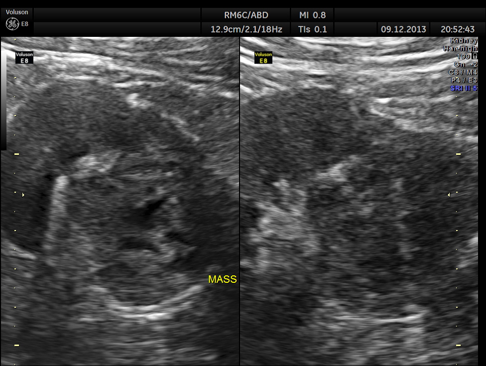
Gray scale reconstruction of the mass is given below.
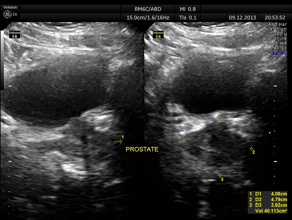
Urinary bladder and prostate are shown below. Mild prostatomegaly seen.
The findings were confirmed with CT Scan . he underwent radical left nephrectomy and the biopsy was reported as RENAL CELL CARCINOMA.
The images presented above show certain features better seen with reconstruction . But the diagnosis as is usual is made out with the 2 D images .

Excellent case of academic interest. Thanks for Post.
LikeLike
Thanks sir
LikeLike
this is a very trained eye. Thank you for sharing these images! very good for me to review!
LikeLike
Thanks
LikeLike
thank u. very informative
LikeLike
Excellent work.
I just forward it to every Sonograher I know.
Thank you!
LikeLike
Thanks sir
LikeLike
Thanks, Kriznan
LikeLike
Pingback: RENAL MASS – HYPERNEPHROMA – RENAL CELL CARCINOMA | Looking Through a Transducer