This was a 55 year old lady who had earlier undergone hernia repair surgery few years ago ,following caesarian section done many years ago.
She came with history of localised pain to the left of the umbilicus for the past 18 days.
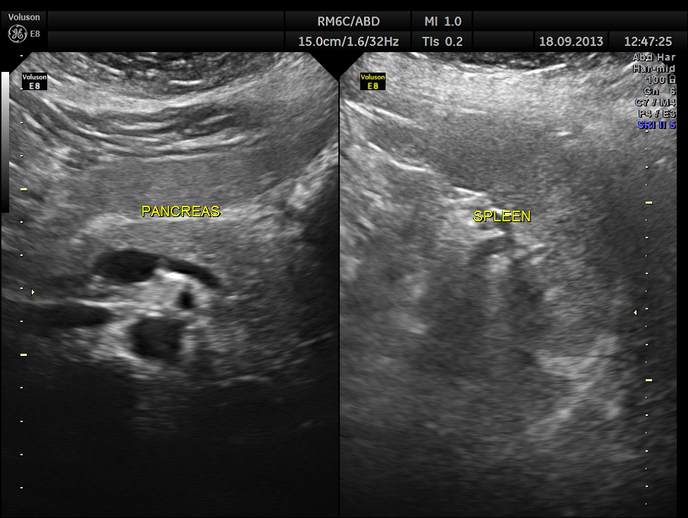
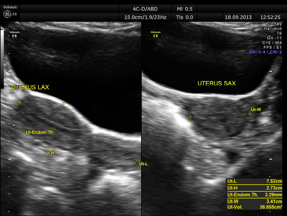
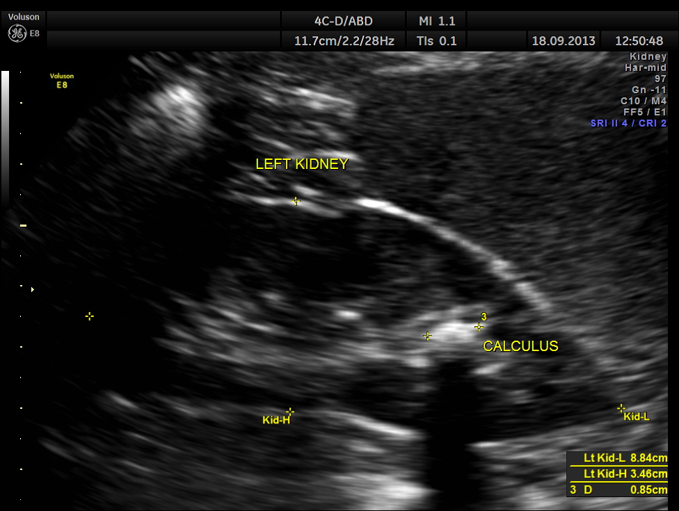
Ultrasound revealed normal liver, gall bladder, pancreas, spleen ,post menopausal shrunk uterus and normal right kidney.

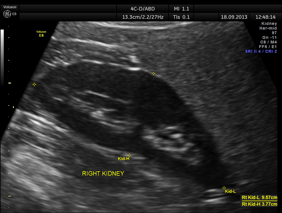
The left kidney showed a calculus.
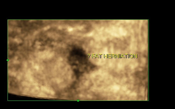
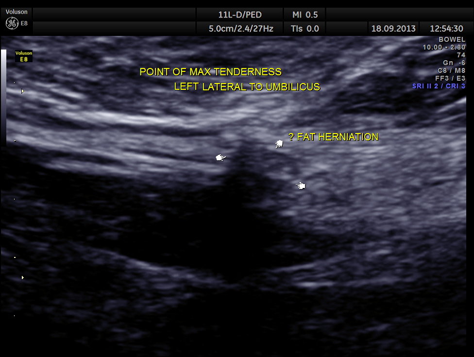
Scan with high resolution probe over the point of maximal tenderness revealed peritoneal fat herniation.
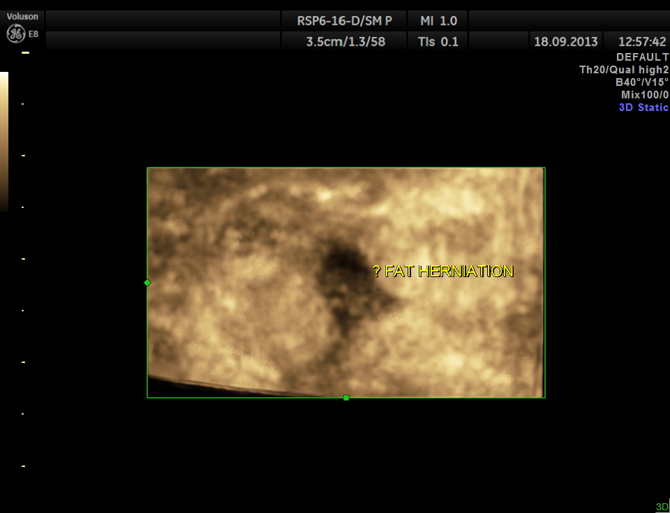
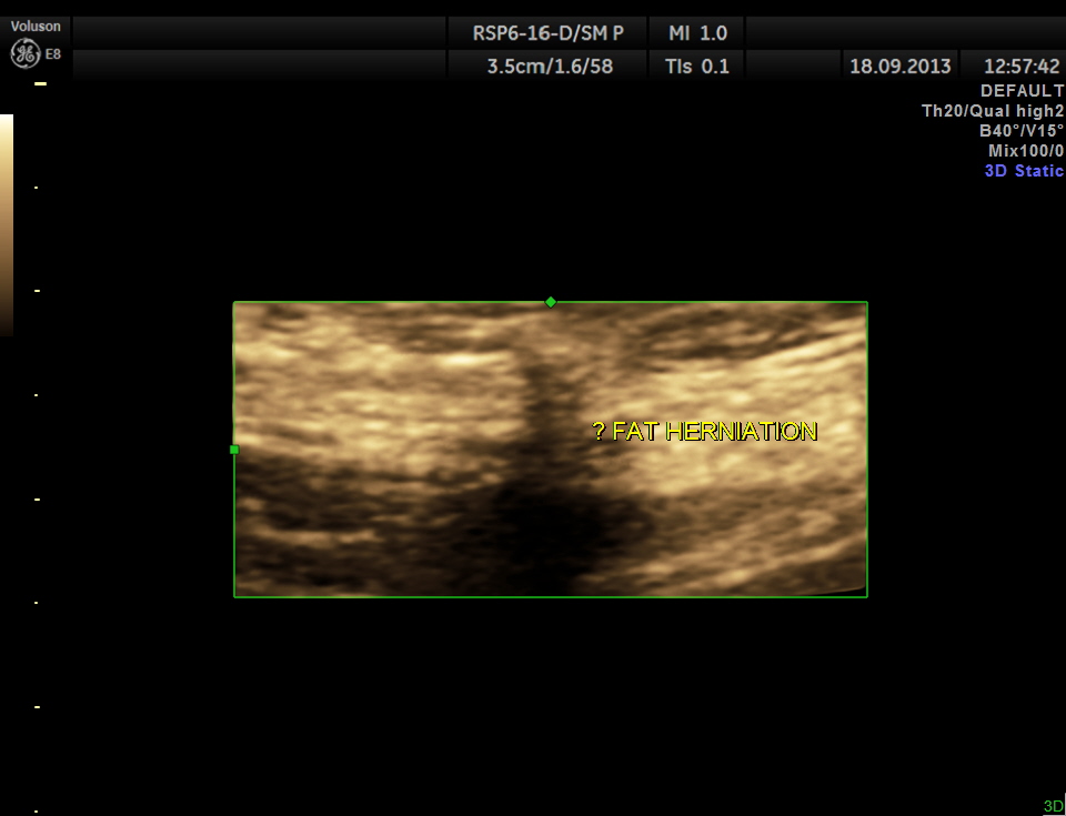
the next image is a 3 D reconstruction.
On specific questionig the patient said that 18 days ago she had to lift and move heavy objects in the house hold ( as her daughter was getting married ) !
the following link takes you to a nice pictorial review of CT images of hernia.
http://radiographics.rsna.org/content/25/6/1501/F34.expansion.html
the following link is a good discussion of ultrasound of the anterior abdominal wall hernias

This is a job well done.
LikeLike
Thanks
LikeLike
Interesting and educational,well done!
LikeLike
Thanks
LikeLike
Thank you for the photos, explanations and SlideShare, a little more I can add insight about the hernia.
LikeLike
Well presented and Thanks for sharing!
LikeLike
Well presented and thanks for sharing.
LikeLike
This is quite informative. Thank you
LikeLike