This was a 57-year-old gentleman who was referred for evaluation of severe pain in the left flank region . He came a couple of days later from the time his physician advised the scan . As he removed his shirt , I found that he had herpes zoster eruptions on the side of the pain . I actually told him that he would not require the scan now . Then he said that he has a dull ache on his right loin region on and off . So I proceeded with the scan .
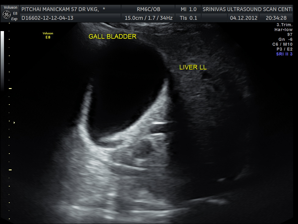
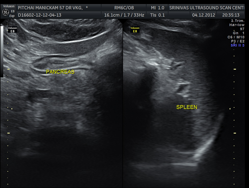
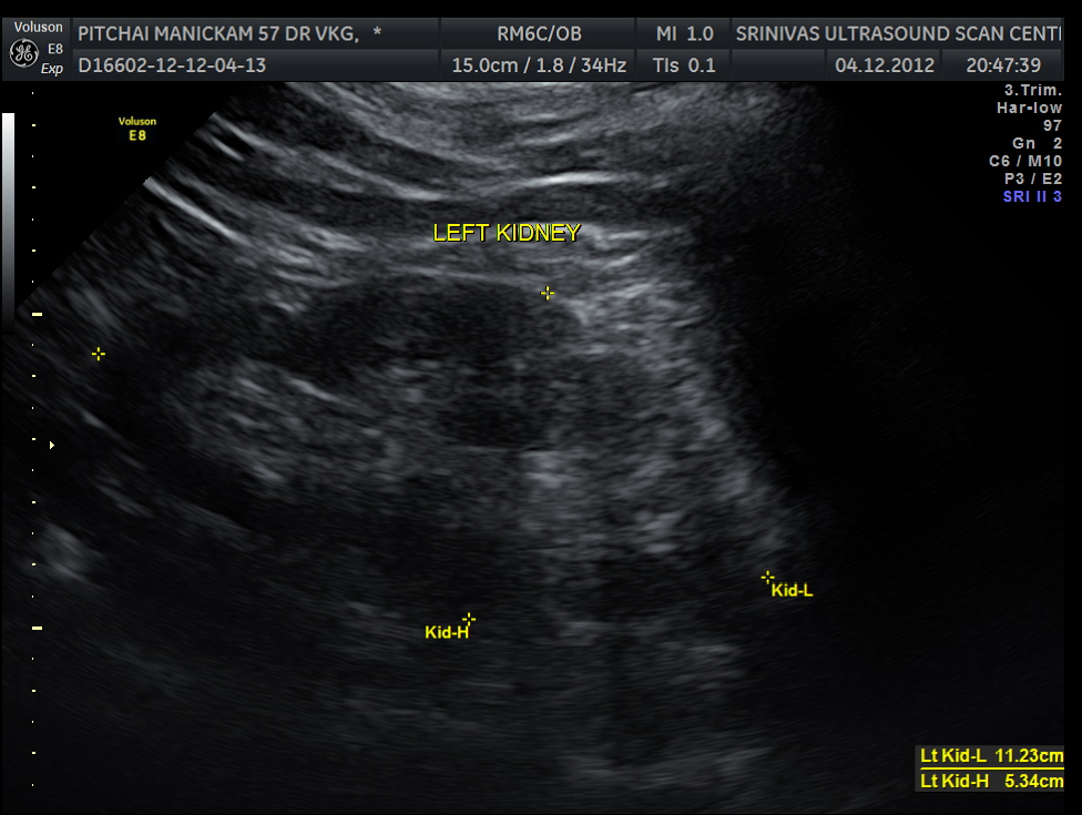
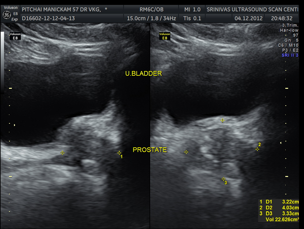
Liver, gall bladder , pancreas, spleen and the left kidney were normal . The prostate showed mild enlargement.
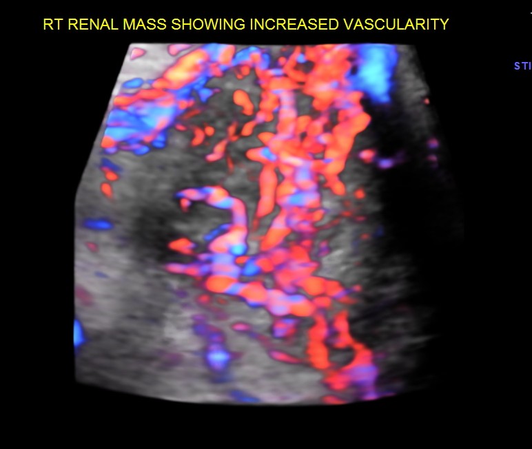
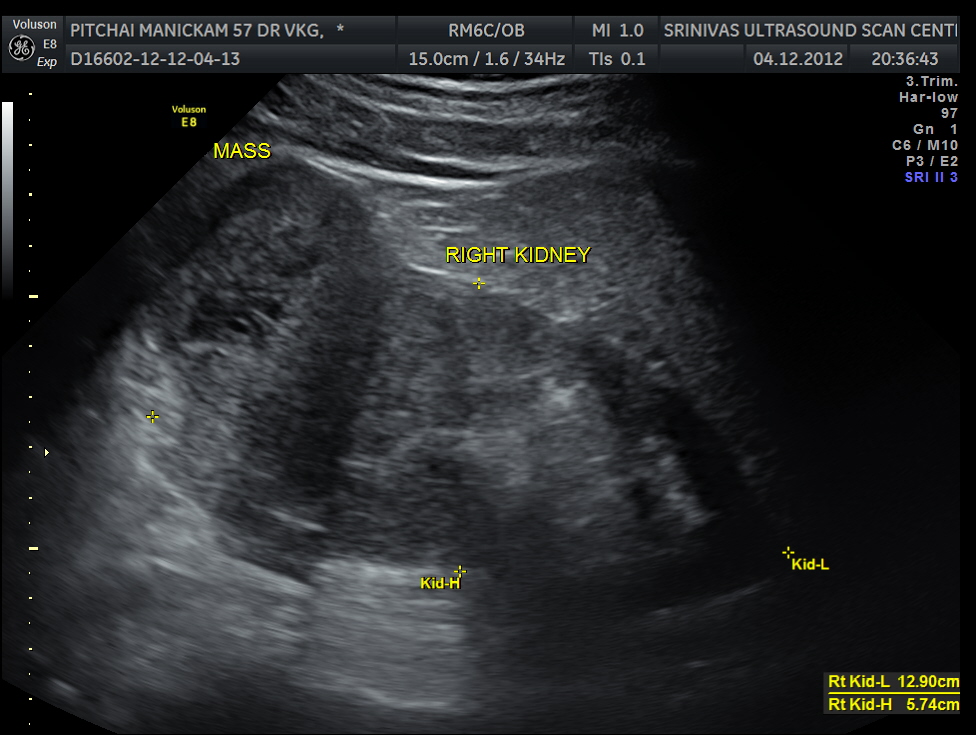
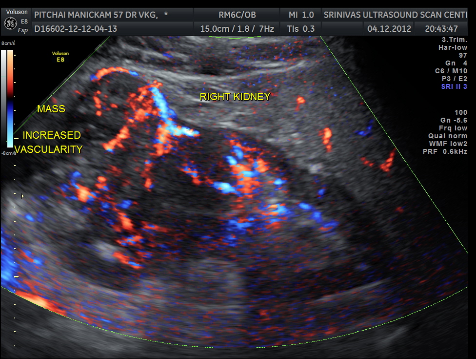
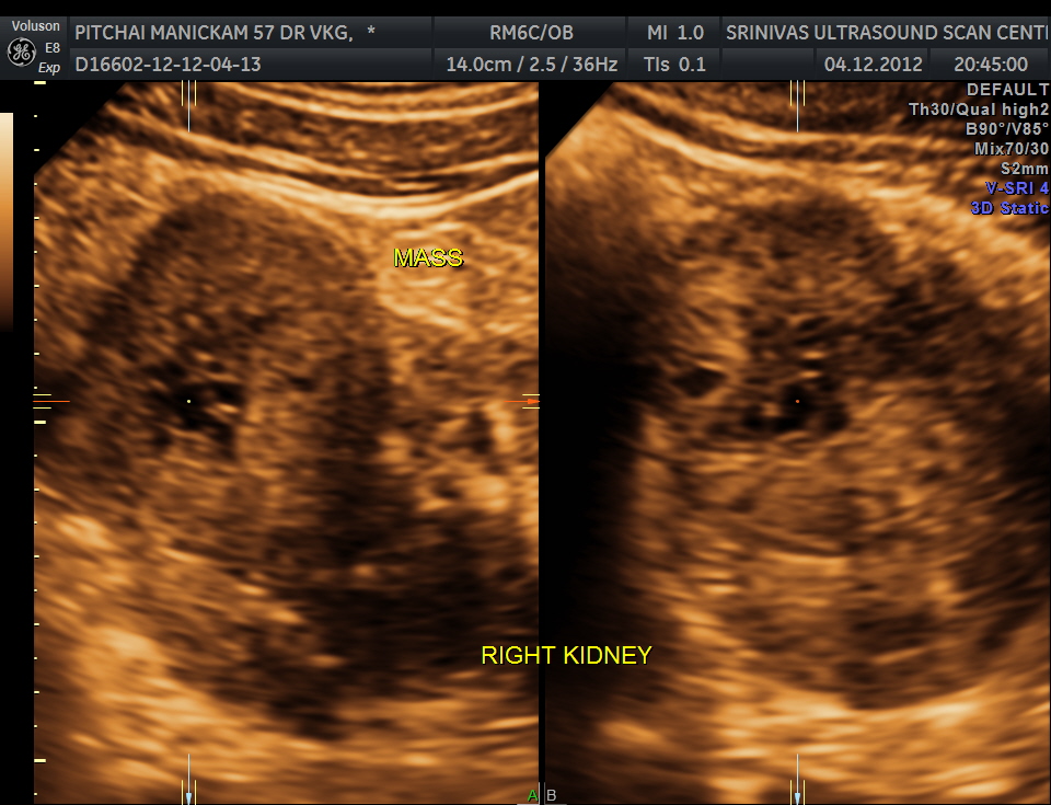
The right kidney pictures are given below. Mass lesion seen in the lower pole .
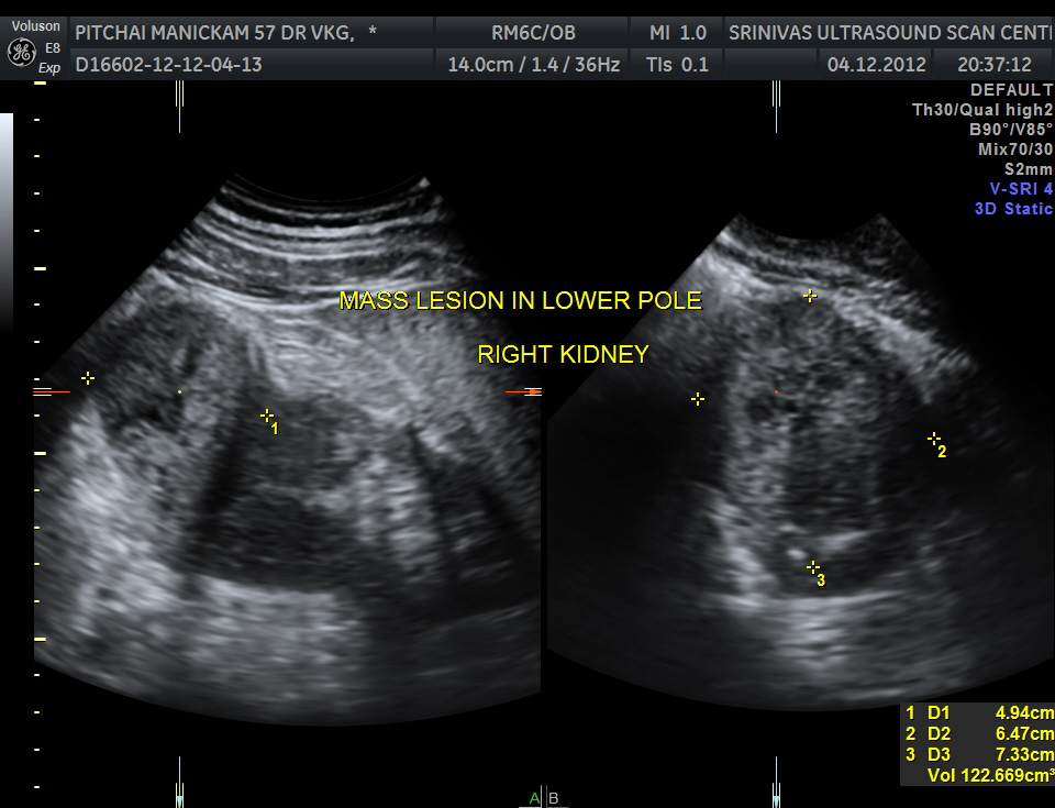
the mass has a volume of around 122 cc
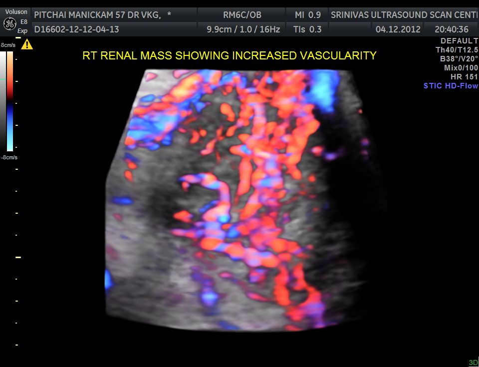
The right renal mass shows increased vascularity .
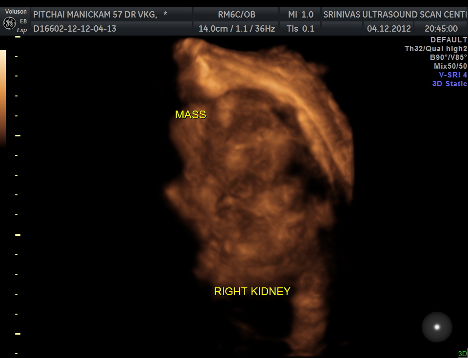
3 D images are given below.
the next is a reconstructed image of the mass.
The ultrasound diagnosis was a right renal mass – likely to be hypernephroma.
The images are presented to show the increased vascularity on colour Doppler and the lobularity seen in the reconstructed image and also for the clinical presentation of pain on the contralateral side due to herpes zoster.
Unfortunately the patient was lost for follow up .

Pingback: RENAL MASS – HYPERNEPHROMA – RENAL CELL CARCINOMA | Looking Through a Transducer