This was 26 year old lady, primigravida, without history of consanguinity.
Scan was done at 32 weeks of gestation. Earlier scans have been reported as normal.
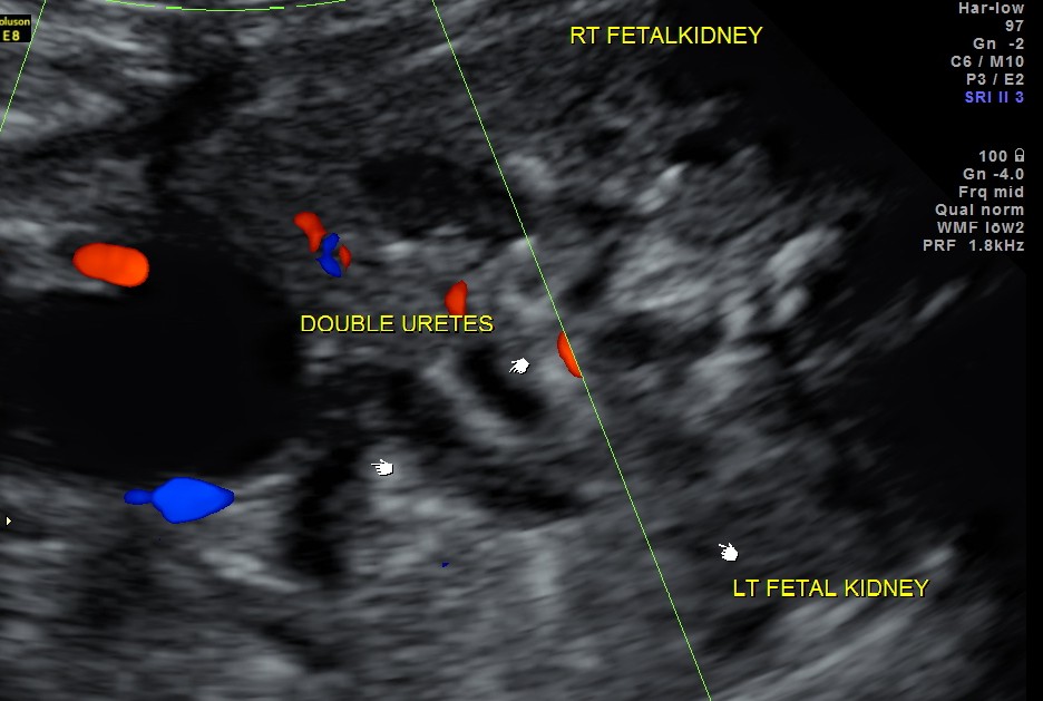
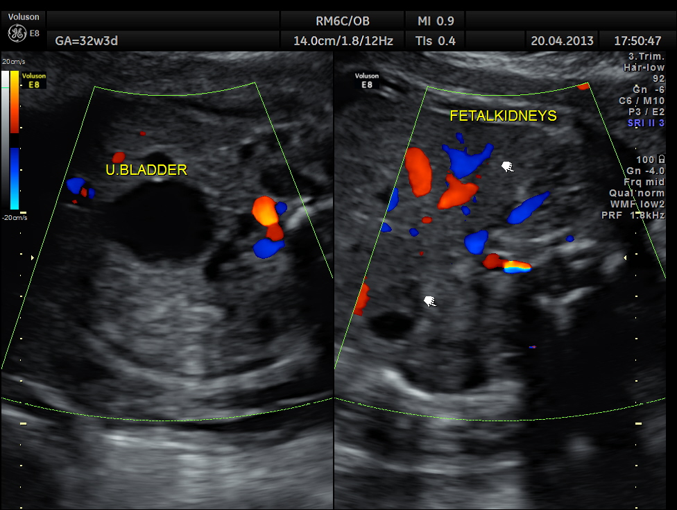
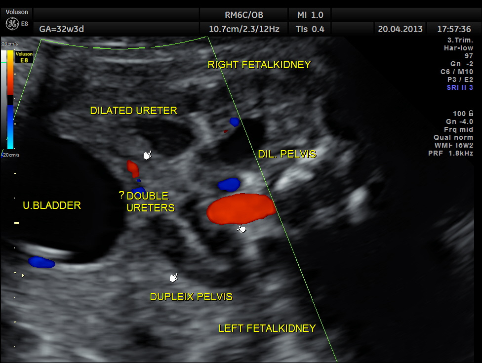
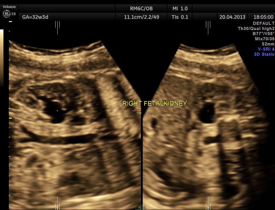
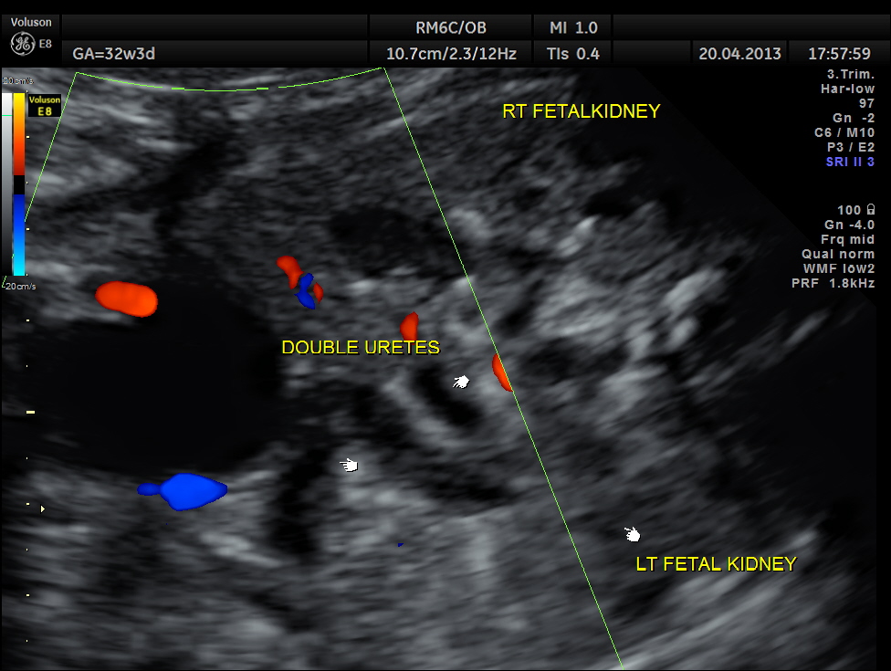
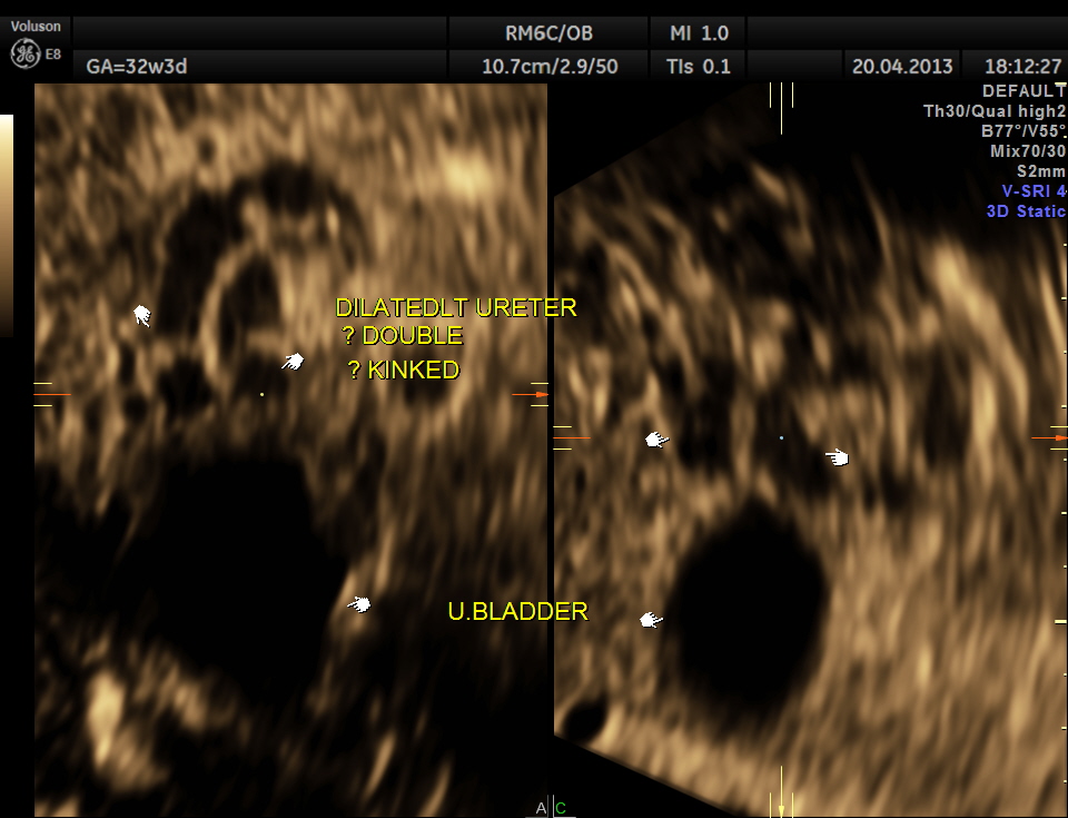
The following images show bilateral renal pelvi ectasis and bilateral dilated ureters with all the changes more in the left kidney .
The left kidney also showed possible duple-ix pelvis with double ureters arising from them . But this finding could not be confirmed in all the views .
The patient was advised to come for a review scan after a month and counselled about the need for procedural correction after delivery.
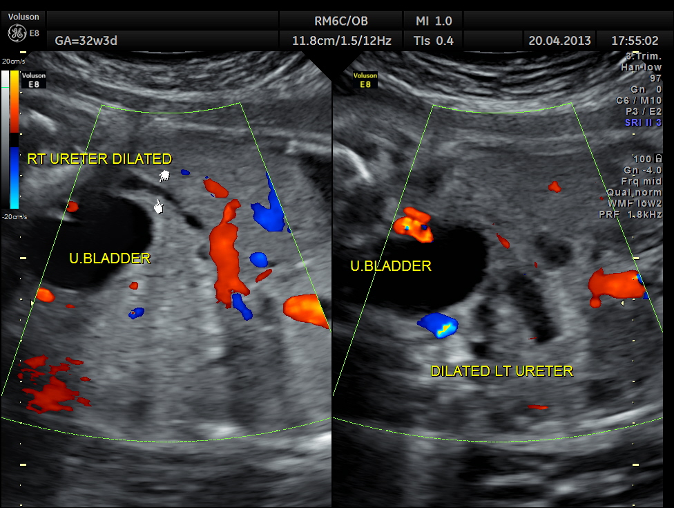
dilated ureters are seen
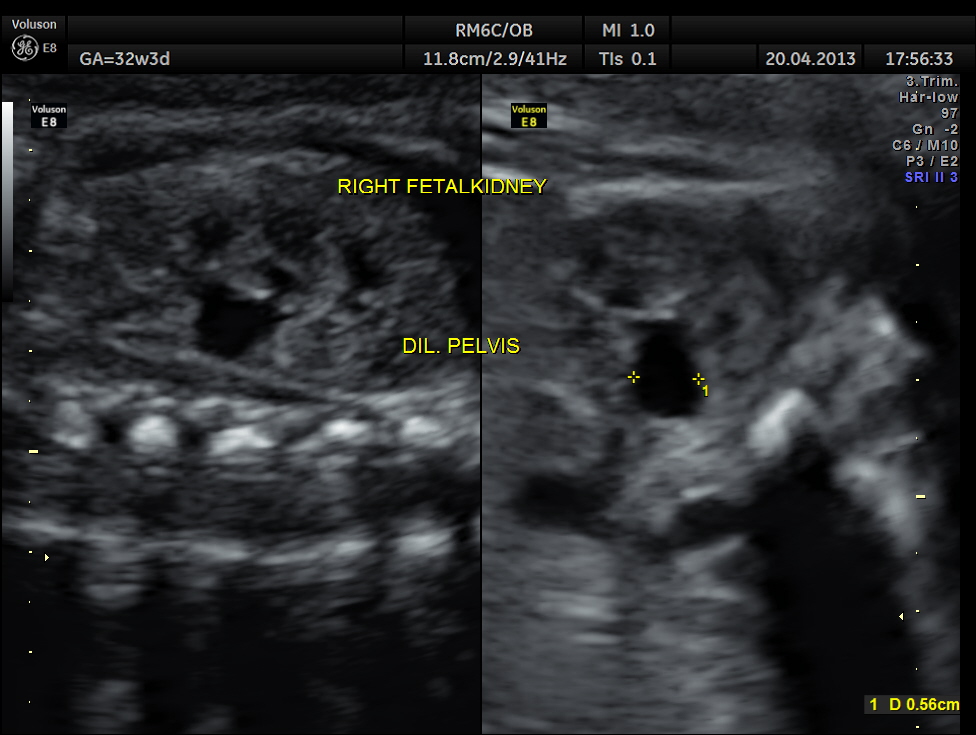
rt renal dilated pelvis
? tortuous and kinked left ureter
also looks like double ureters
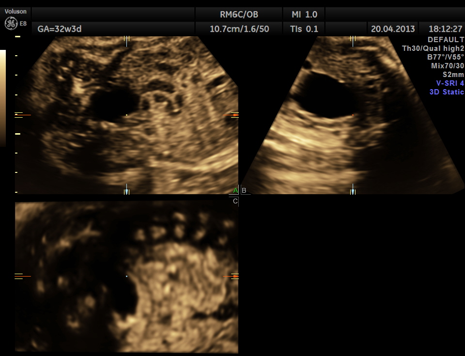
3 d image

is it a case of posterior urethral valve
LikeLike
Axial images of kidney would better showed the urethral dilation
LikeLike