This was a 19 year old primi with history of consanguinity.The scan was done in the first trimester.
A large cystic mass was made out in the lower abdomen.

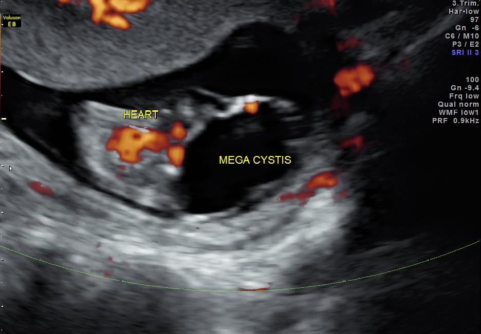
MULTI PLANAR VIEW OF FETUS SHOWING GROSSLY ENLARGED BLADDER


The following text about mega cystis can be found in http://radiopaedia.org/articles/fetal-megacystis.
Fetal megacystis refers to the presence of an unusually large bladder in a fetus. It is generally defined as a
- bladder diameter > 7 mm in the first trimester 3
- bladder diameter > 30 mm in the second trimester
- bladder diameter > 60 mm in the third trimester
Epidemiology
The estimated incidence of antenatal imaging is at ~ 1 : 1500 pregnancies.
Pathology
It can result from a number of causes but with the main underlying mechanism being either a distal stenosis of reflux.
Associations
Associated anomalies are common 6 and include
- posterior urethral valves
- chromosomal anomalies
- on a first trimester scan ( 10 – 14 weeks)
- if the longitudinal bladder diameter of 7 – 15 mm there is a risk of a chromosomal defects is esimated at ~ 25% 4
- if the bladder diameter is > 15 mm the risk of chromosomal defects is estimated at ~ 10% 4
- on a first trimester scan ( 10 – 14 weeks)
- oligohydramnios
- megacystis microcolon intestinal hypoperistalsis (MMIH) syndrome (Berdon syndrome)
- megacystis megaureter syndrome
- prune belly syndrome
-
Radiographic assessment
Antenatal ultrasound
Will show an enlarged bladder
Ancilliary sonographic findings
- may show evidence of oligohydramnios
- may show associated renal anomalies
Treatment and prognosis
The overall prognosis can be variable from progressive obstruction to spontaneous resolution. A follow-up ultrasound is necessary to correctly interpret the significance of megacystis detected in the first trimester
If the fetus is chromosmally normal and there is megacystis on the 1st trimester scan
- there is spontaneous resolution of the megacystis in about 90% of cases when the 1sttrimester longitudinal bladder diameter is between 7 – 15 mm 4.
- if the bladder diameter is > 15 mm there is a very high likelihood of associated with progressive obstructive uropathy 4
Management will depend on the underlying pathology

I’ve never seen a picture of this before. Good work!
LikeLike
Thanks
LikeLike
very good
LikeLike