Please note that the ultrasound pictures of this patient are from our records. The PET CT images are done elsewhere at Chennai. They are reproduced here as they give the full picture of the clinical presentation.
This 74 year old gentleman has been coming to me for more than 10 years for mild systemic hypertension and hypothyroidism.He used to smoke cigars until a few years ago. 3 years ago he developed unexplained giddiness and on evaluation was found to have pituitary macroadenoma. Endocrine and neuro surgical consulatations were sought and he was on cabergoline and was doing very well. In April 2012 , he came for a routine review and was asked to continue his existing medications . One week later he came again with severe right upper quadrant pain and aversion to food. There was no history of cough in the recent past. He was advised an ultrasound scan of the abdomen and the findings are as below.

heterogenous liver texture

multiple irregular nodules seen

nodules with altered texture
CT scan of the abdomen confirmed the findings ; For further clarification PET whole body scan was done at a centre in Chennai. The following images are from that.

Heterogeneously enhancing irregular spiculated soft tissue mass measuring ~ 34 x 29 mm noted in the apical segment of right upper lobe. The lesion abuts the pleura posteriorly.
FDG avid enhancing lymph nodes noted in right and left paratracheal regions, largest measuring ~ 19 x 11 mm on the right side.

lung mass

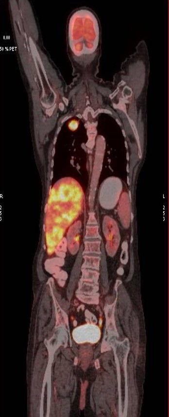
LIVER SECONDARIES

PET CT IMAGE OF LIVER SECONDARIES

D10 SPINE LESION


The whole body PET CT
The final diagnosis was
- Metabolically active spiculated right upper lobe lung mass – likely malignant primary.
Mediastinal and retroperitoneal FDG avid metastatic lymphadenopathy.
Extensive FDG avid hepatic metastases involving both lobes.
Focal FDG avid lesion in left lamina of D10 – possibly metastatic.
The biopsy report of the liver secondary was a poorly differentiated carcinoma.
The oncologist advised palliative care and the patient passed away 3 weeks later.
The important lesson from this presentation was that the patient presented with silent lung Ca with extensive secondaries in a short duration of time.

Thanks for sharing this. Malignancy, always a masquerader . Needs a high index of suspicion.
LikeLike
Thanks great case, how do u differentiate primary hepatic masses from secondary
LikeLike
Very impressive. Thank You
LikeLike
Some oncologists prescribe sorafenib, but there are many side-effects.
LikeLike