
This was a 24 year old primi with history of consanguinity sent for evaluation of probable dextrocardia.
The following images were seen .
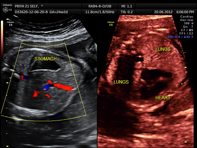
the stomach bubble is on the left ; the heart is on the right side
the following video shows right atrium and rt ventricle to be grossly dilated and the left atrium and left ventricle to be very small and hypoplastic.
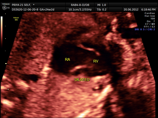
dilated RA AND RV with hypoplastic left heart
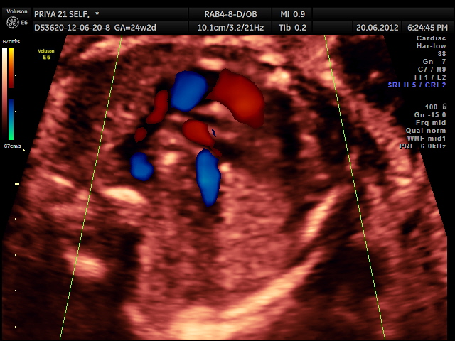
colour flow shows dilated RA and RV with hypoplastic LA and LV
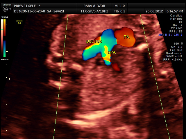
RV outflow tract with dilated Pulmonary trunk dividing into the Rt Pulm artery and the ductus seen. Aorta is not made out. ; SVC is seen
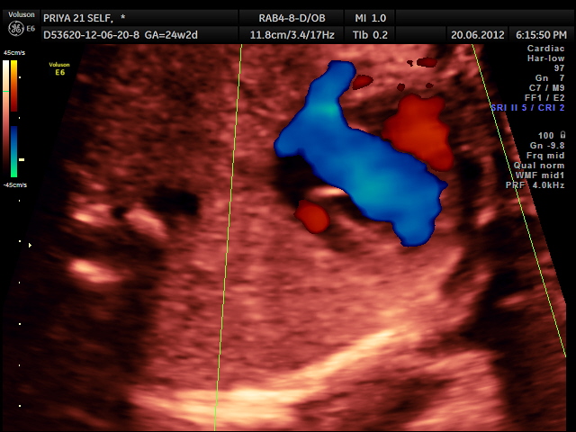
another view of RVOT , PA with the divisions ; Aortic flow not made out clearly
The excerpts given below are from the following website :http://www.chop.edu/service/cardiac-center/heart-conditions/hypoplastic-left-heart-syndrome-hlhs.html
Hypoplastic left heart syndrome (HLHS) is a severe congenital heart defect in which the left side of the heart is underdeveloped.
The heart’s left side has the job of pumping oxygenated blood into the aorta, the large artery that carries blood to the body. In a child with HLHS:
- The mitral valve, which separates the two left chambers of the heart, is too small or completely closed (atretic).
- The left ventricle (the lower, pumping chamber) is very small.
- The aortic valve, which separates the left ventricle and the aorta, is too small or completely closed (atretic).
-
What are the treatment options?
Hypoplastic left heart syndrome (HLHS) is most often fatal without early intervention. It will typically require open heart surgery to re-direct the oxygen-rich (“red”) blood and oxygen-poor (“blue”) blood in a series of three reconstructive operations known as “Staged Reconstruction.”
-
Stage I – Norwood procedure -occurs within a few days of birth.
-
Stage II – Glenn procedure – occurs within four to six months of birth
-
Stage III – Fontan procedure -occurs between 1 1/2 to 4 years of age.
