Looking Through a Transducer

Kriznan's ultrasound images

Menu

Skip to content
  • Home
  • About

VESICAL CALCULI

Date: June 11, 2012Author: kriznan Category: Abdominal Ultrasound, Genito-urinary Tags: 3 D RECONSTRUCTED IMAGE OF VESICAL CALCULI, BLADDER STONES, URINARY BLADDER CALCULI, VESICAL CALCULI

Post navigation

← SUPRASPINATUS TEAR
URINARY BLADDER CARCINOMA →

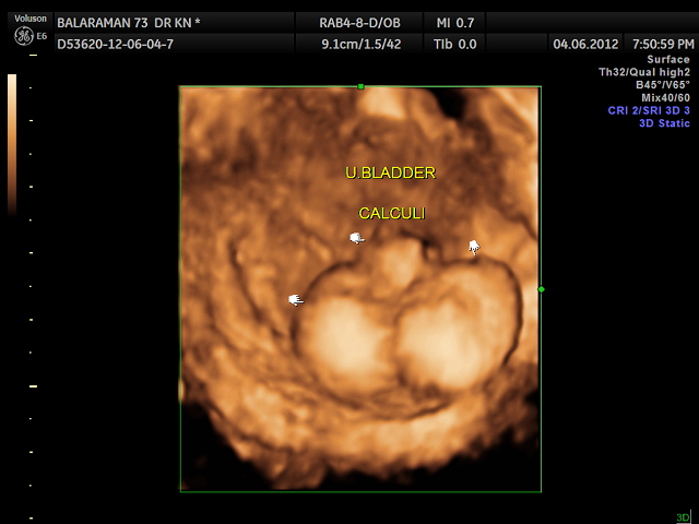
THIS WAS A 73 YEAR OLD MAN WITH DIFFICULTY IN PASSING URINE AND STRANGURY OF RECENT ONSET ; THE REFERRING PHYSICIAN WAS KEEN ON RULING OUT ANY PROSTATIC PATHOLOGY.

USG SHOWED THIS

This slideshow requires JavaScript.

USG THROWS UP SURPRISES !

Share this:

  • Click to share on X (Opens in new window) X
  • Click to share on Facebook (Opens in new window) Facebook
  • Click to email a link to a friend (Opens in new window) Email
  • Click to share on LinkedIn (Opens in new window) LinkedIn
  • Click to share on WhatsApp (Opens in new window) WhatsApp
  • More
  • Click to print (Opens in new window) Print
  • Click to share on Reddit (Opens in new window) Reddit
  • Click to share on Tumblr (Opens in new window) Tumblr
  • Click to share on Pinterest (Opens in new window) Pinterest
  • Click to share on Pocket (Opens in new window) Pocket
  • Click to share on Telegram (Opens in new window) Telegram
Like Loading...

Blog Stats

  • 240,059 hits

Leave a comment Cancel reply

Recent Posts

  • Mass Lesion in 3rd Ventricle October 8, 2017
  • D.O.R.V. December 4, 2016
  • What is your diagnosis? July 27, 2016
  • CONJOINED TWINS June 12, 2016
  • Rt aortic arch and aberrant left subclavian artery May 20, 2016

Archives

Enter your email address to follow this blog and receive notifications of new posts by email.

Join 1,315 other subscribers

Like my page on Facebook

Like my page on Facebook

Top Posts & Pages

  • Appendicular mass
  • COMPLEX HYDROCELE
  • LIMB BODY WALL COMPLEX OR BODY STALK COMPLEX
  • Bladder calculus & Prostatomegaly - 2D & 3 D images
  • PVD -PERIPHERAL VASCULAR DISEASE
  • UNEXPLAINED ANEMIA IN A YOUNG MAN
  • OBSTRUCTIVE HYDROCEPHALUS AND AMNIOTIC BAND ( 3D )
  • IUGR - PLACENTOMEGALY - ABNORMAL DOPPLER - UTERO PLACENTAL INSUFFICIENCY
  • Agenesis of Corpus Callosum
  • VARICOCELE

Categories

  • 3 d images
  • A V MALFORMATION
  • Abdominal Ultrasound
  • anemia
  • appendix
  • BILE DUCT
  • bowel
  • colour doppler
  • DOPPLER
  • Echo cardiography
  • Fetal Echocardiography
  • fetal face
  • fetal neuro-sonography
  • fetal spine
  • Fetal Ultrasound
  • Gall bladder
  • GASTRO-INTESTINAL
  • Genito-urinary
  • GENITO-URINARY
  • Gynaecology
  • hereditary spherocytosis
  • IUGR
  • jaundice
  • KIDNEYS
  • Liver
  • LOWER LIMB ARTERIES
  • Musculo skeletal ultrasound
  • NEURO SONOGRAPHY
  • OBSTETRIC
  • Pancreas
  • PLACENTA
  • PORTAL VEIN
  • Scrotal ultrasound
  • Shoulder, Musculo-skeletal ultrasound
  • Spleen
  • THYROID
  • TWINS
  • ultrasound images
  • Uncategorized
  • urinary bladder
  • Uterine arterio venous malformation
  • VASCULAR
Blog at WordPress.com.
  • Comment
  • Reblog
  • Subscribe Subscribed
    • Looking Through a Transducer
    • Join 195 other subscribers
    • Already have a WordPress.com account? Log in now.
    • Looking Through a Transducer
    • Subscribe Subscribed
    • Sign up
    • Log in
    • Copy shortlink
    • Report this content
    • View post in Reader
    • Manage subscriptions
    • Collapse this bar
%d