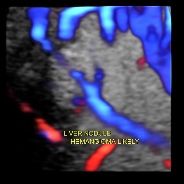
Hemangioma of the liver is generally an innocuous incidental finding . At times the differential diagnosis considered would include a solitary secondary nodule. This would require further imaging investigations like CT and MRI. Colour and Power Doppler could be helpful in demonstrating the feeding vessel to a hemangioma , but is very difficult to demonstrate at times. 3D 4 D glass body reconstruction could help in demonstrating the feeding vessel.
This was a 60 year old gentleman who was being evaluated for dyspepsia.
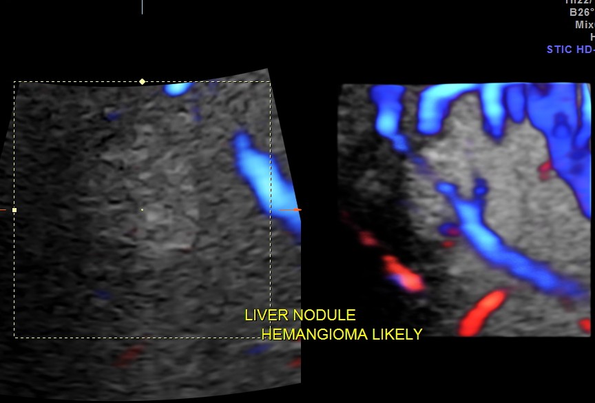

The pictures and the video are presented to show the clarity with which glass body reconstruction demonstrates the feeding vessel .
some references are given below.
http://www.nlm.nih.gov/medlineplus/ency/article/000243.htm
http://radiopaedia.org/articles/hepatic-haemangioma

very much instructive
LikeLike
very interesting i enjoyed reading about this and the doppler pictures were very clear. thanks
LikeLike
Very good, thanks for these information !
LikeLike