This was a 42 year old man with left scrotal swelling , with history of pain on standing and lifting weights.
Clinically the surgeon felt that this could be varicocele. The patient had undergone repair surgery for left inguinal hernia few years ago.
The following images were obtained .
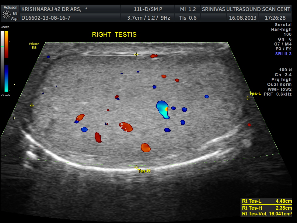
rt testis cc
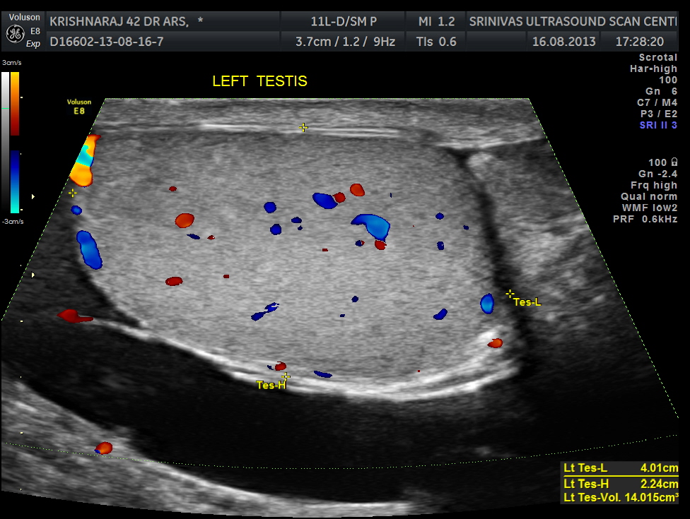
LEFT TESTIS ; HYDROCELE SEEN
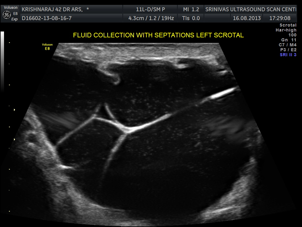
SEPTATED HYDROCELE WITHINTERNAL ECHOES SEEN

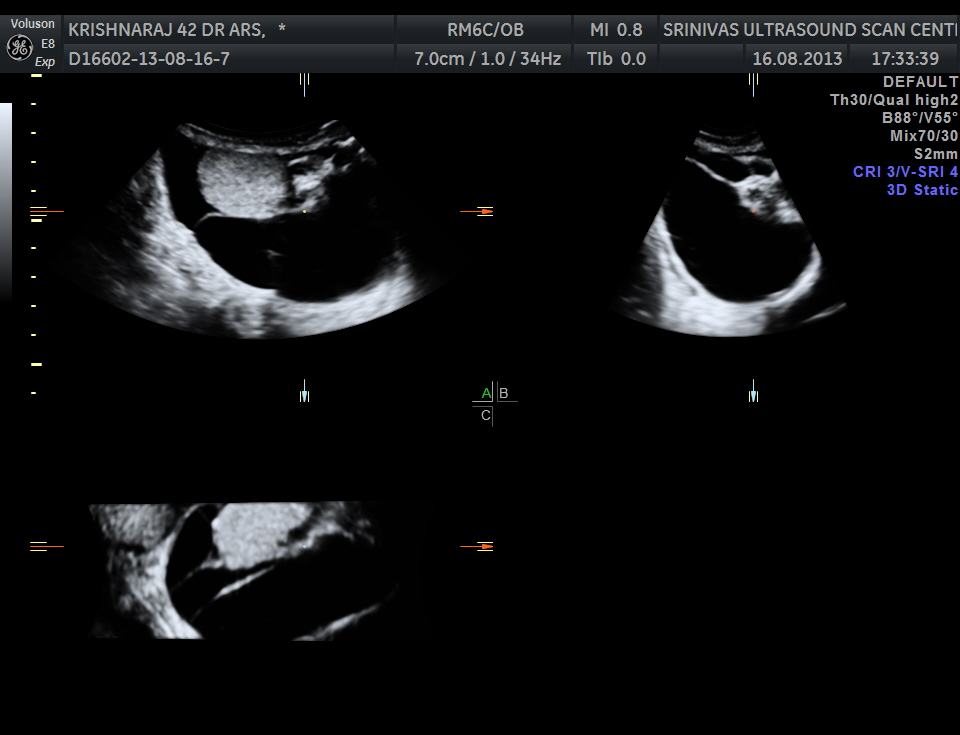
MULTI PLANAR VIEW OF SEPTATED HYDROCELE
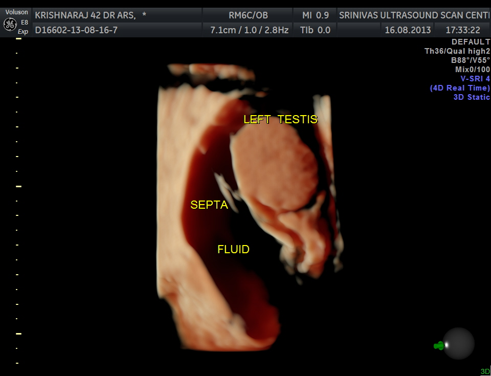
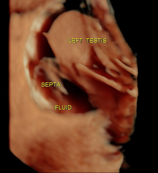
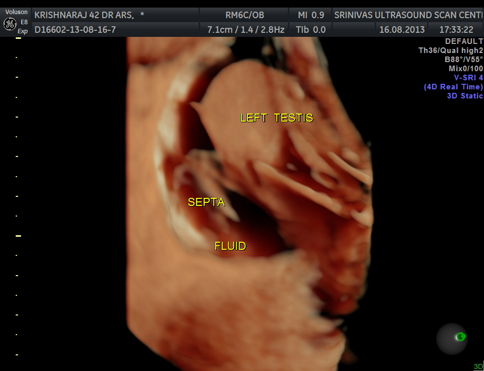
3 D HIGH DEFINITION VIEW OF THE SAME
In general most surgeons do not refer patients for a scan when they are clinically very sure of hydrocele. In this patient the surgeon’s clinical impression was varicocele and so he wanted a scan .
The finding of hydrocele is helpful before surgery. But does the diagnosis of a septated hydrocele change anything for the surgeon ?
When the fluid contains high protein or cholesterol content, the hydrocele may appear complex or septated. Hematoceles and pyoceles are complex hydroceles. Sonographically, a simple hydrocele is seen as an anechoic dark fluid collection surrounding the testicle , whereas a complex hydrocele may contain internal echoes with septations and loculations.
Probably for the surgeon , there is no difference in dealing with a simple or complex hydrocele .

Fantastic images, The 3D scans are impressive as always.
LikeLike
Thanks
LikeLike
I’ve never used the 3D function before…now Im really interested in using it!! Great pics!
LikeLike
Try it ; it is a lot of fun
LikeLike
As usual, very educational images! well done!
LikeLike
ive never used 3d b4 but looking at these images 2D still does it for me,
LikeLike
More than reconstruction , multi planar imaging is helpful
LikeLike
Great images!
LikeLike
great images!
LikeLike
Filaria should be ruled out.
LikeLike
We should be cautious about scrotal malignancy also.
LikeLike