
This was a 22 year old lady referred for evaluation of polyhydramnios.
She had severe polyhydramnios.

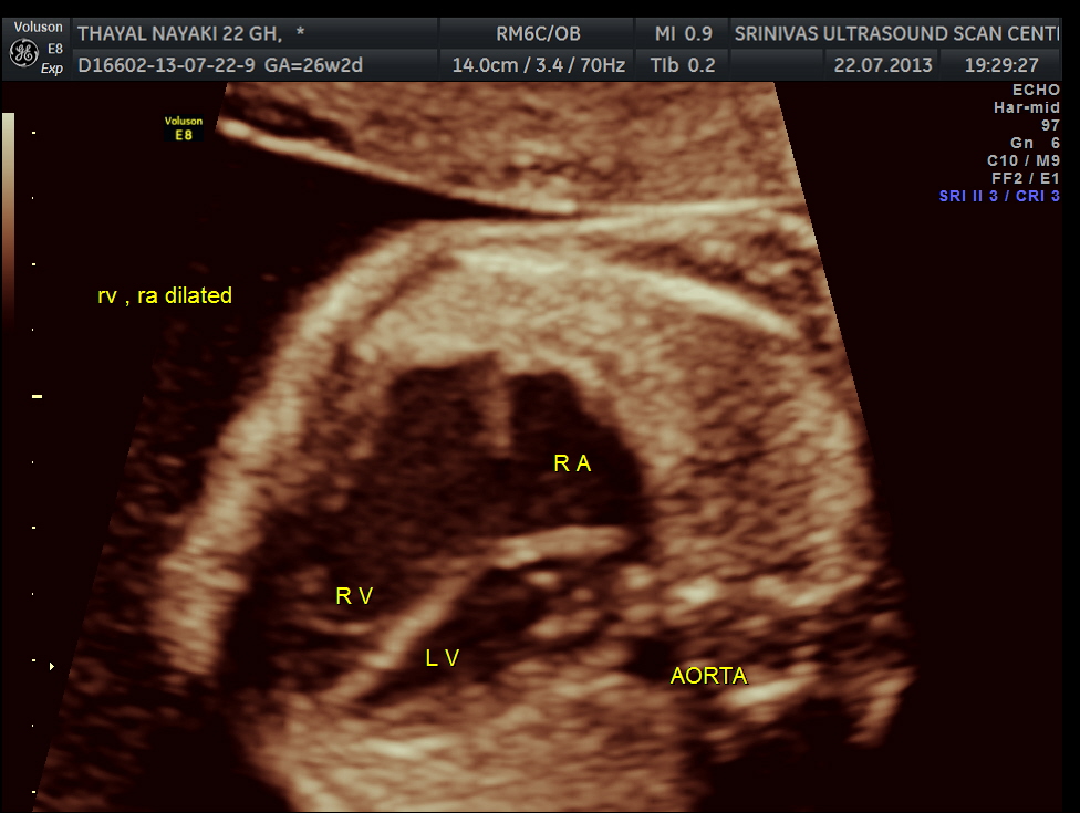
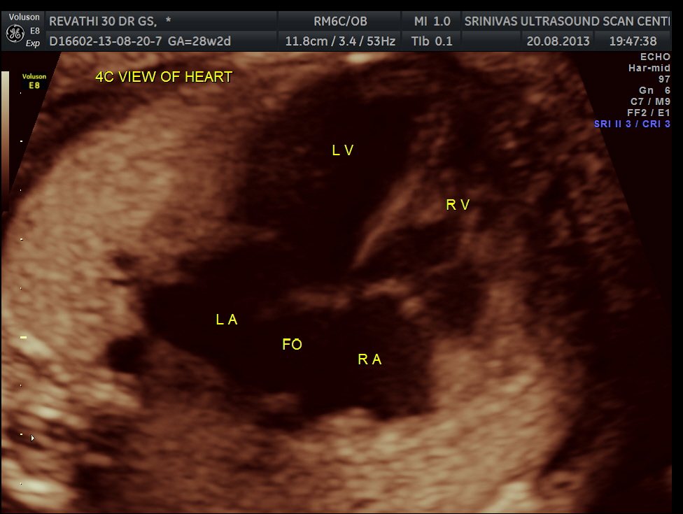
The cardiac axis appeared to be abnormal .
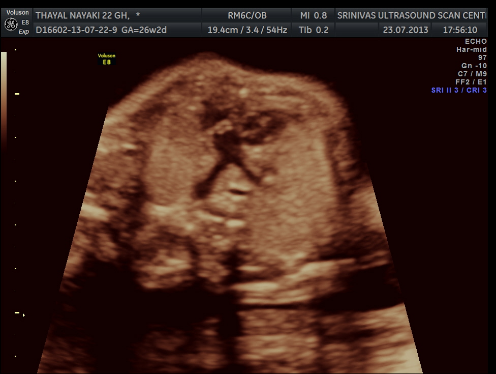
4 chamber view is given below .
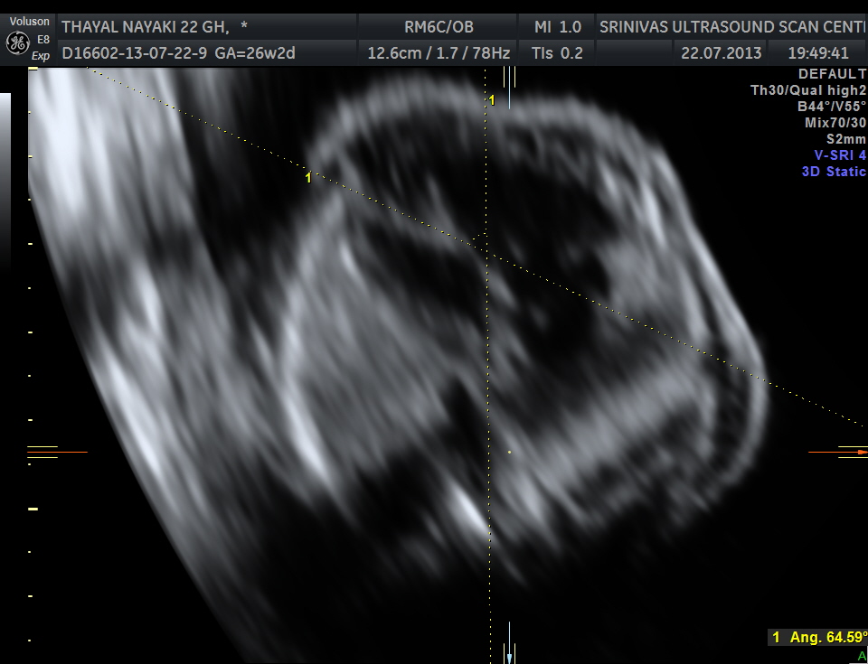
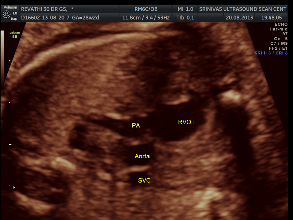
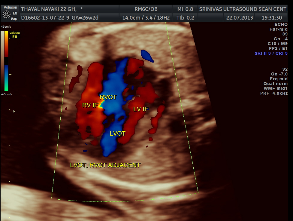
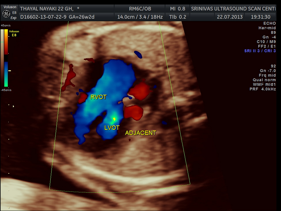
3 vessel view shows a prominent single vessel , which is very suggestive of outflow tract anomalies like transposition of great arteries, double outlet right ventricle , truncus arteriosus and corrected transposition of great arteries.
another view of the above two
Under normal circumstances the chamber in front of descending aorta is left atrium ; and the aorta arises from the left ventricle which has no trabeculations ; the pumonary artery which divides into the two branches arises from the anterior right ventricle and crosses over the aorta .
some normal images are given below to compare with the latter images.
normal 4 chamber view
next is the normal outflow tracts and the normal 3 vessel view and the normal arches.
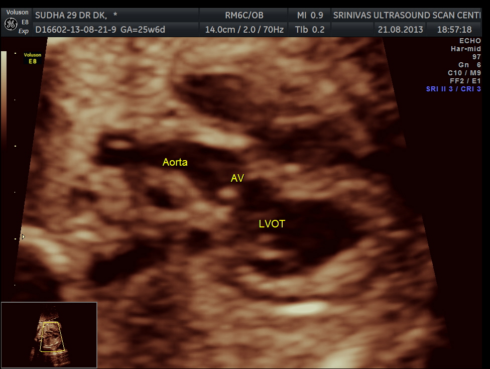
normal lvot giving rise to the aorta
normal 3 vessel view
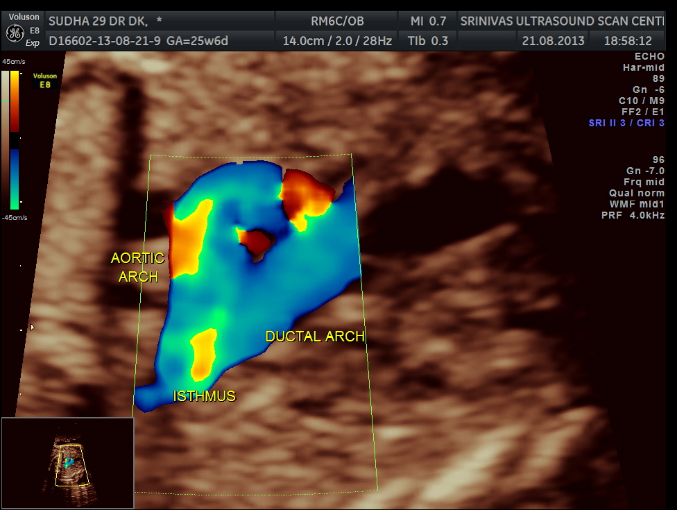
ductal and aortic arches
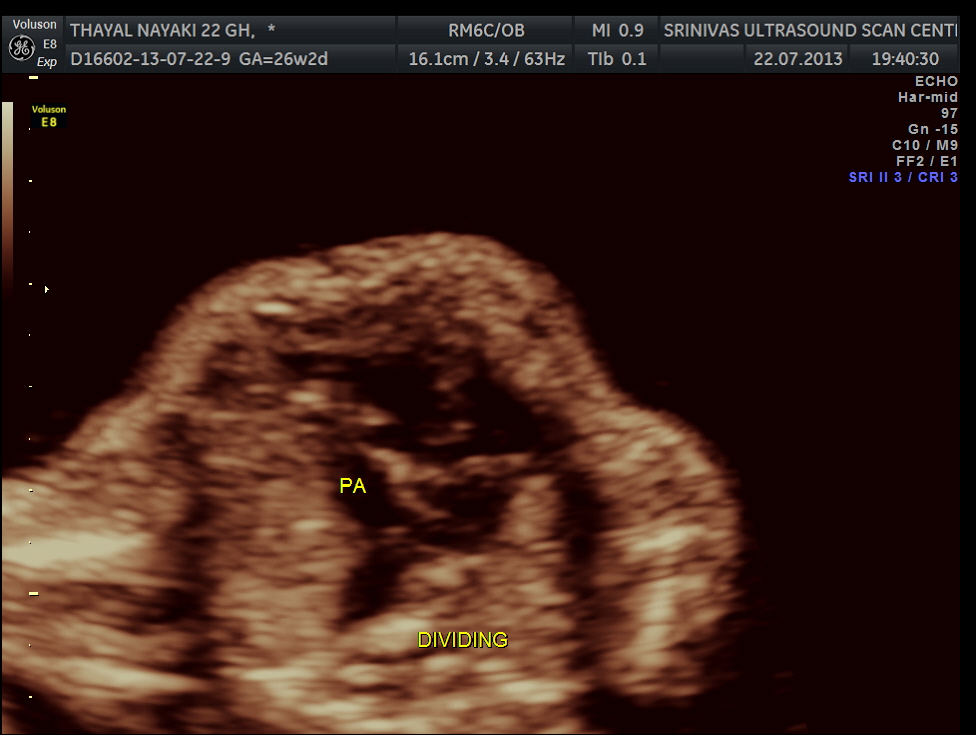
here we can see the pulmonary artery with its two branches arising from the lower (left) ventricle .and the aorta arising from the anterior (right ) ventricle.
aa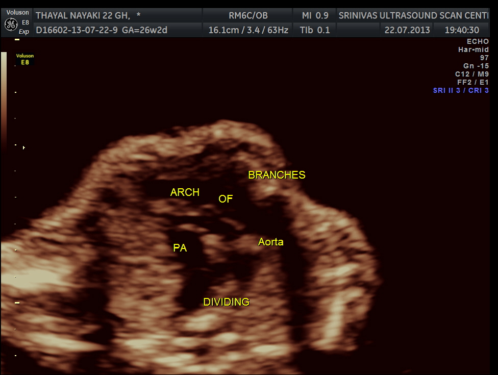
pulmonary artery dividing into two
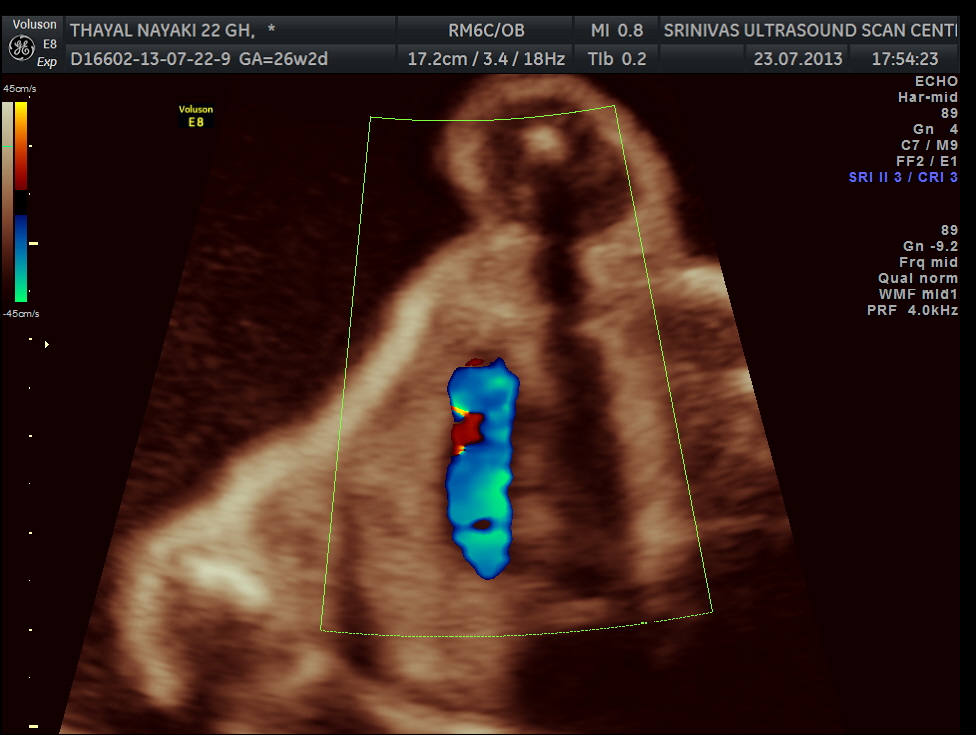
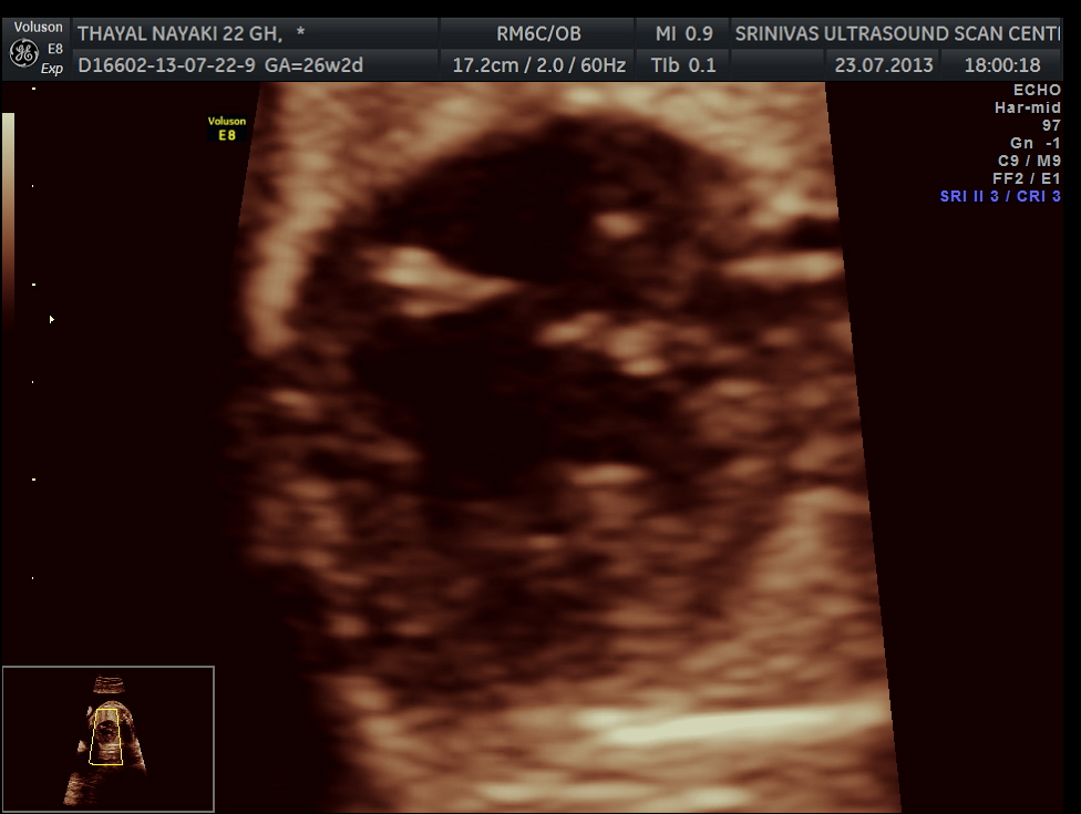
vsd is seen in the picture below.
generally colour flow imaging is not said to be very helpful
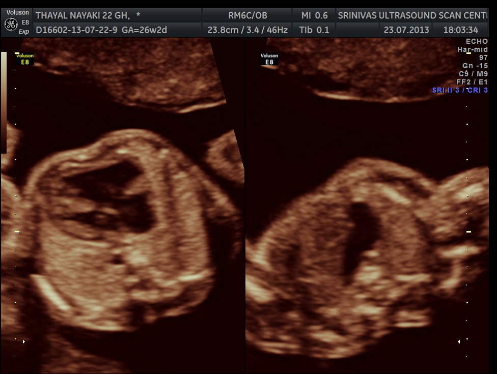
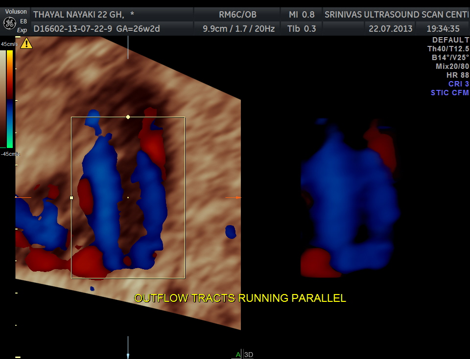
the following 3 D reconstructed image shows the parallel flow of the great arteries
THE DIAGNOSIS OFFERED WAS TRANSPOSITION OF GREAT ARTERIES WITH VENTRICULAR SEPTAL DEFECT
Transposition of the great arteries (TGA) is the most common cyanotic congenital heart lesion that presents in neonates. The hallmark of transposition of the great arteries is ventriculoarterial discordance, in which the aorta arises from the morphologic right ventricle and the pulmonary artery arises from the morphologic left ventricle.
Factors in the mother that may increase the risk of this condition include:
- Age over 40
- Alcoholism
- Diabetes
- Poor nutrition during pregnancy (prenatal nutrition)
Our patient did not have any of the above.
TGA has 4 major types and knowing them earlier would help in the neonatal management.
the following links offer more information.
http://emedicine.medscape.com/article/900574

Has she been asymptomatic for 22 yrs?
LikeLike
This was an obstetric scan ; the mother was 22 years old ; the findings described are for the foetus
LikeLike
We also had TGA here of patient with no real risk factors; Takes you by surprise…
Nice case.
LikeLike
very good case & excellent images .amazing .
LikeLike
Thanks
LikeLike
Good job, clear images. Please share with as most interesting cases!
LikeLike