sorry for the long gap in posting a case .
the last case was vesical calculi ; this one was a urinary bladder carcinoma .
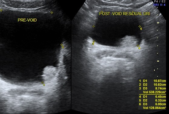
47 year old man presented with complaints of painless hematuria ; he had a similar self limiting episode 3 months ago , which he ignored.His ultrasound pictures are given below

an irregular mass lesion seen in the lateral wall
 the next is a 3 d reconstruction , which shows the prominent median lobe prominence adjacent to the mass.


the median lobe prominence is well appreciated in the 2 d image also.
the next is a post void picture showing the mass

this patient underwent surgery and histopathology proved the diagnosis of bladder carcinoma and he is doing well now.

commendable efforts
LikeLike
Very cool! Thanks for this.
LikeLike