This was a very interesting problem . I learnt a few things over a period of time . May be many others would have got the diagnosis easily.
This was a 27 year old man working as an I.T.consultant in a city. He started getting headaches , which were disabling . The headache was mostly one sided . He was evaluated for the headache ; His CT scan of the brain was normal. He had features of iron defeciency anemia with thrombocytosis . His neurologist after a bit of trial and error made a diagnosis of ‘ indomethacin sensitive chronic paroxysmal hemicranial headache ‘ . He responded very well to indomethacin . But his anemia was not improving with medical treatment.
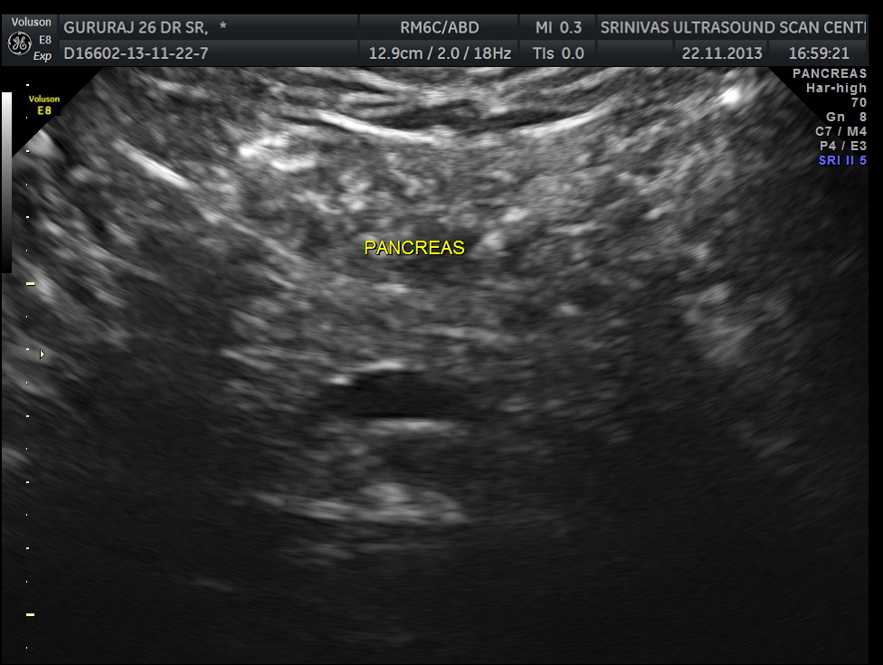
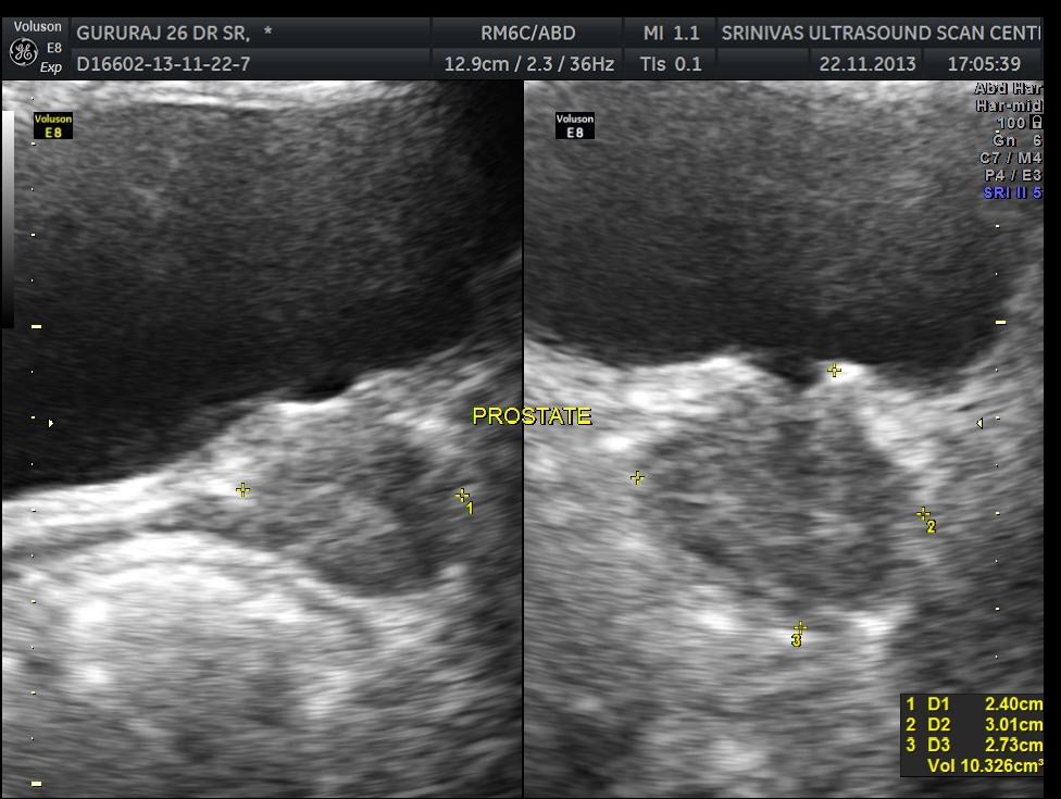
An ultrasound abdomen was done as part of evaluation of anemia.


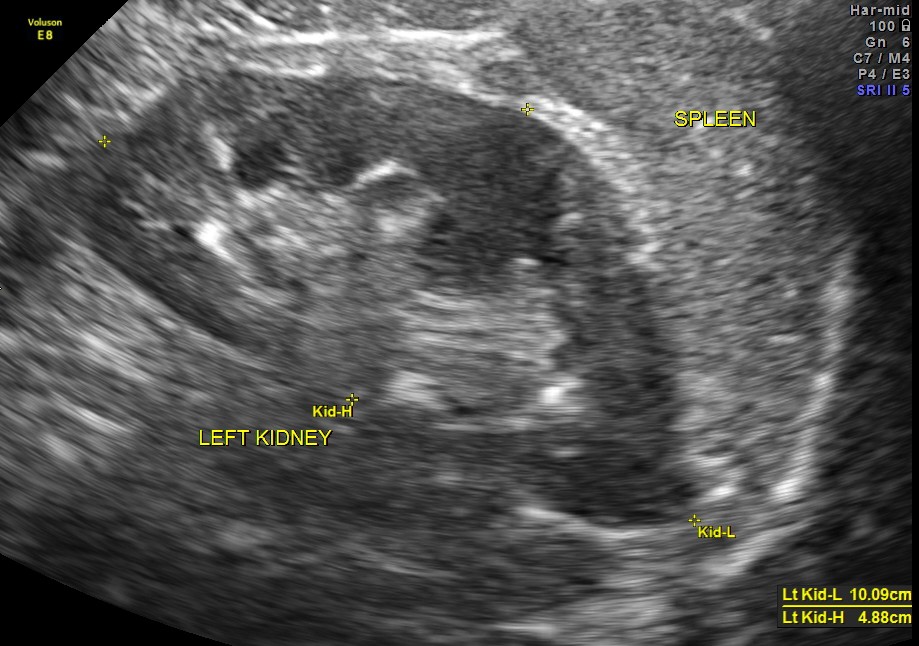
Now for the unexpected findings . To finish off the scan , I was sweeping the aorta, IVC and the pre and post aortic regions.
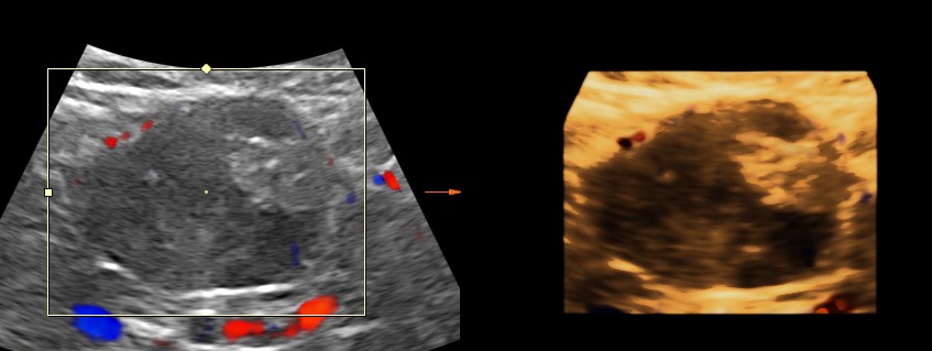
The following image was obtained with the transducer in the mid epigastric region and an inch to the left of the mid line.
This mass visualised there . This was antero-lateral to the aorta and showed mixed echotexture , with some vascularity.
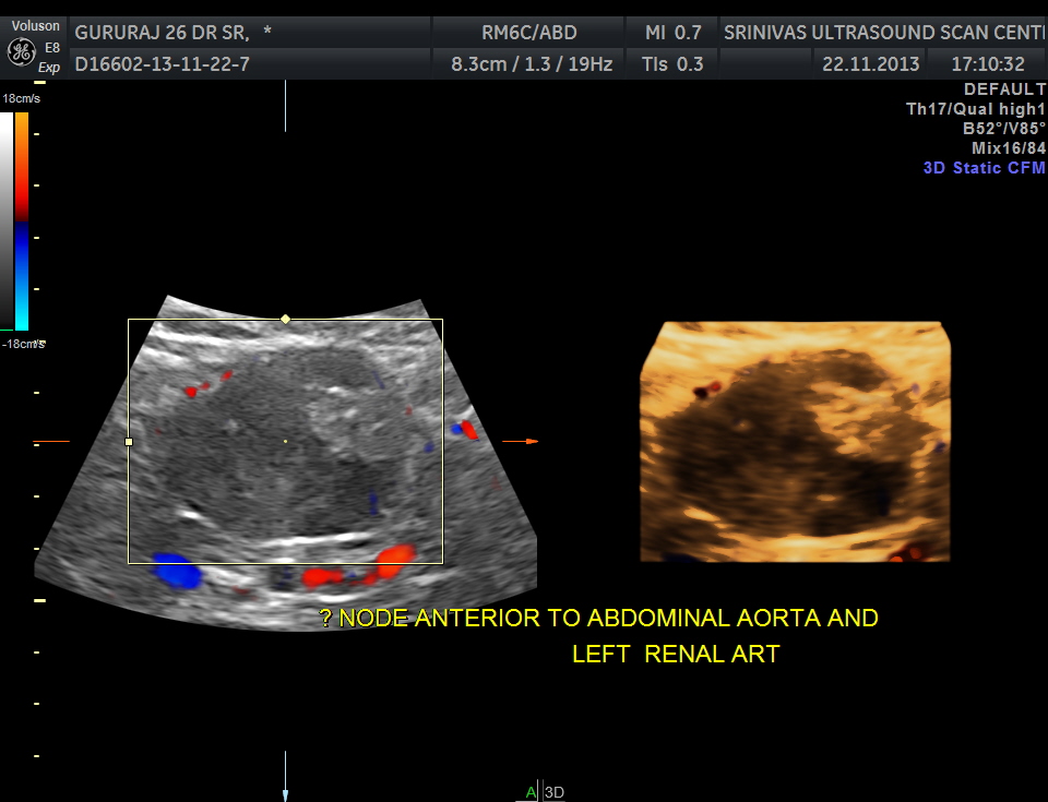

This was not mobile and all i could offer was a description of what was seen with the impression of a mass of unknown origin – ?? enlarged node and advised further work up.
He underwent a contrast CT and was found to have a mass arising from the intestinal wall. He underwent surgery subsequently and the mass was removed . It was a benign leiomyoma arising from the jejunal wall and showed some erosions on the inner aspect ( cause for the anemia ) . Now came the pleasant surprise for everybody.His anemia improved and his headaches disappeared totally. The explanation offered was reactive thrombocytosis due to the anemia probably caused the hemicranial pain and when that was taken care of he was better.
Many others looking at the ultrasound image could have probably guessed the pathology. But I was happy that at least I picked up the mass , which led to the other things.

Very intresting case sir!!!
LikeLike
Thanks DR Kishore
LikeLike
very interesting case, thank you for sharing it.
LikeLike
Thanks
LikeLike
too best
LikeLike
Great job.
LikeLike
Interesting Dr Krish. However there is also a possibility that the tumor could have been releasing some vasoactive neurotransmitters that could have also been the cause for the headache. (sothing like carcinoid- though I am not sure if leiomyoma of intestine do so. gs
LikeLike
Hi gs, thanks for your comments. As you say why the headache disappeared is speculative.
LikeLike
Thanks for the interesting case! Great info!
LikeLike
MY CONGRATS FOR VERY NICE AND EDUCATIVE CASE.
DID’NT YOU TAKE IMAGES WITH LINEAR PROBE TOO?
LikeLike
Dear Kriz, I request you to publish it in a journal. you get the ct pictures & also the HPE slide photo.It will be accepted in any international journal.
LikeLike
WELL DONE
GOOD HOME WORK
KEEP IT UP
BUT THAT’S KRISH ALWAYS
LikeLike
Very interesting case. Thank you!
I only have a case of gastric leiomyoma
LikeLike
Thank you very much for presenting this unusual case in a very nice way. I also have learned some new things.
LikeLike
This must have been a thorough scan you did. Well done!
LikeLike
That was pretty awesome thanks you did a great work!
LikeLike
Good study to share. I got some great informations!!!!
LikeLike
Thanks
LikeLike
Awesome case. Its amazing how your instant just guide you to sweep threw the aorta . Amazing case.
LikeLike
Amazing case. Thanks for sharing
LikeLike
Could it be that the tumor did produce serotonin too (like in carcinoid syndrome) causing cefalea then disappeared?
LikeLike
Possible
LikeLike
Definitely a theory to ponder about
LikeLike