In general for anybody these terms are frightening and sound a lot confusing .
Is there any way to simplify things ?
The relationship among the inferior vena cava, aorta, and spine displayed by ultrasonography has been shown to be a reliable method to diagnose situs in the newborn. This technique can be applied to the fetus in utero as well.
Normally, the aorta lies to the left of the spine and the inferior vena cava lies to the right. The following image shows the normal situs in a fetus , with head in upper pole.
The diagnosis of situs inversus is made when the mirror image of the normal pattern is seen.
Right isomerism or asplenia is suggested when the aorta and vena cava are found together on the same side of the spine.

juxtaposition of aorta and ivc in a case of right isomerism ( renal dysplasia also seen )
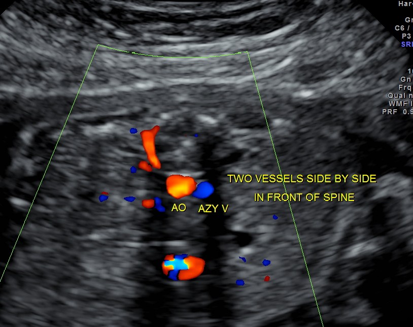
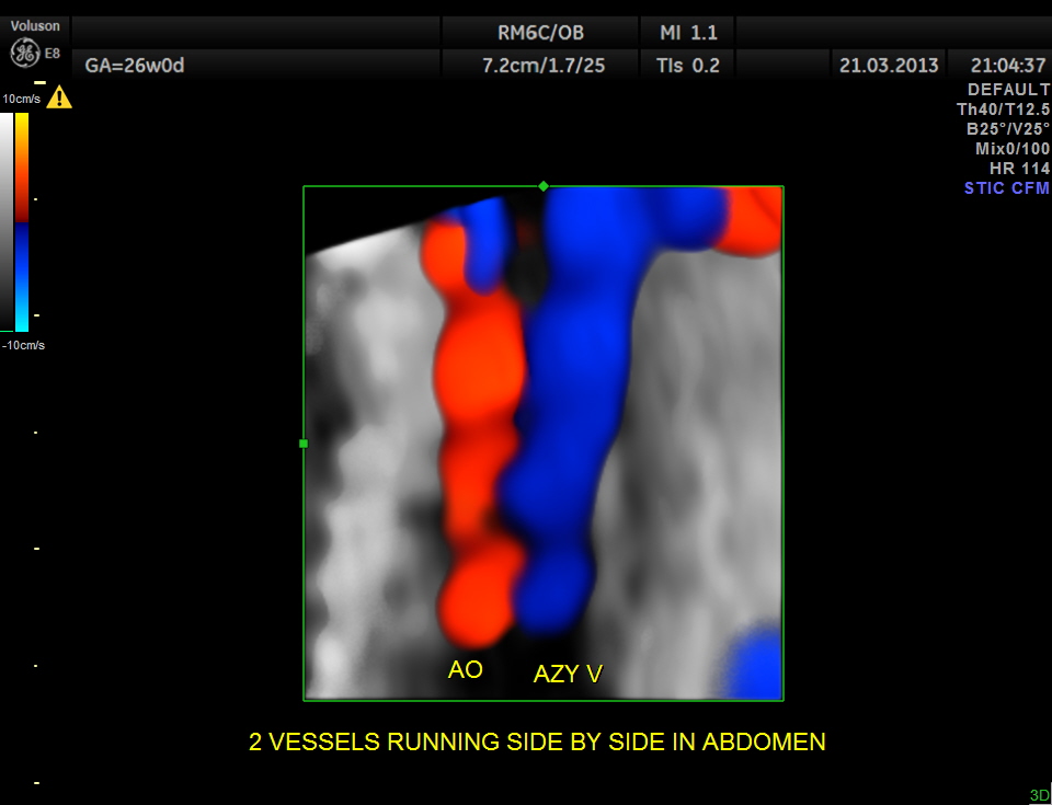
Particular to the diagnosis of Left isomerism or polysplenia syndrome is the demonstration of inferior vena caval interruption with azygos continuation.
The current case is a 29 year old lady with h/o consanguinity ( married her mother’s brother ( uncle).
The fetal lie was : head in upper pole , spine was anterior

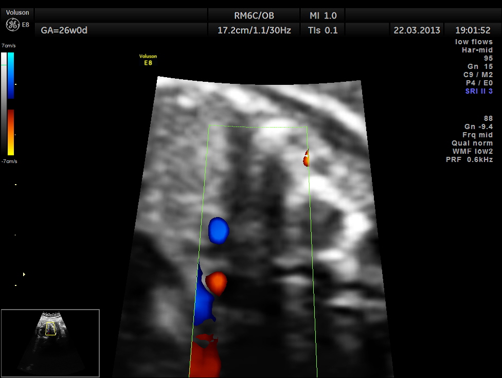
double vessel sign seen in front of spine
two splenic arteries are seen running backwards – indicating polysplenia
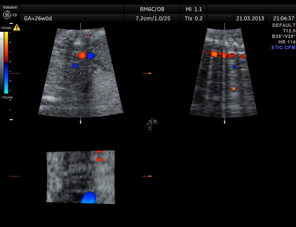
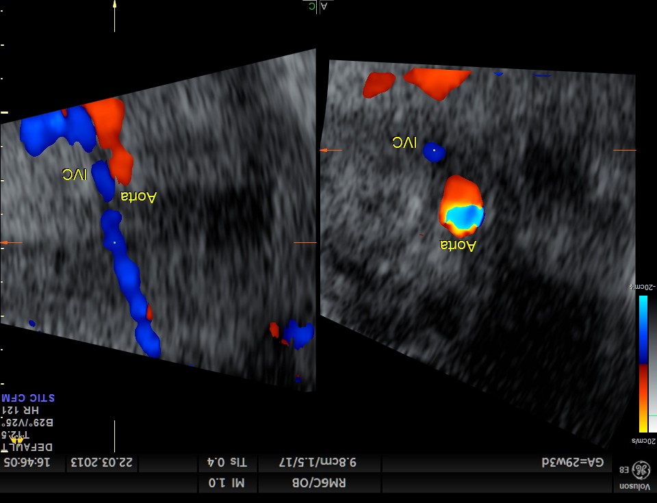
double vessel sign
double vessel sign seen behind the heart
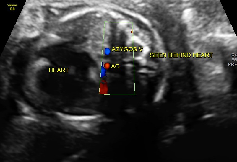
double vessel sign seen behind the heart
zoom of double vessel behind the heart
cardiac axis of less than 29 or more than 59 is considered abnormal ; in this fetus it was 65
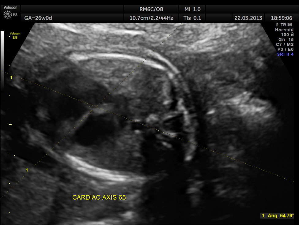
cardiac axis 65
associated abnormalities are bradycardia or complete heart block and atrio ventricular septal defect , commonly unbalanced.
this fetus showed intermittent A.V.DISSOCIATION, BRADYCARDIA AND NORMAL RHYTHM
the following picture shows A.V.DISSOCIATION ( ventricular beats above and atrial below)
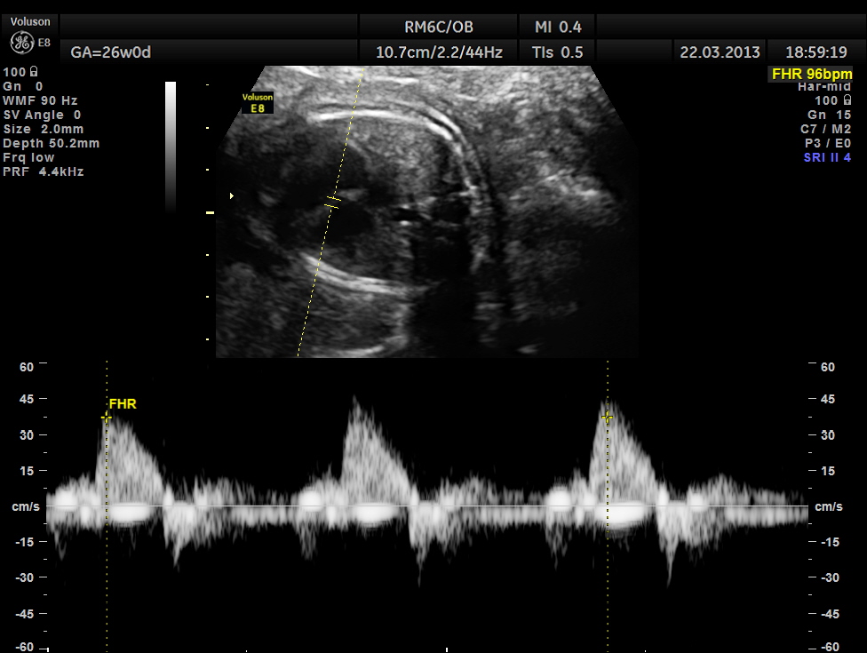
ventricular rate( above mid line ) is 96 bpm ;
atrial rhythm ( seen below base line ) shows dissociation
the following picture shows a probable transmitted rhythm with severe bradycardia
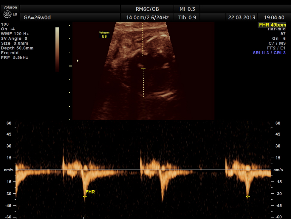
this shows severe bradycardia
the following picture shows a normal conduction and rate
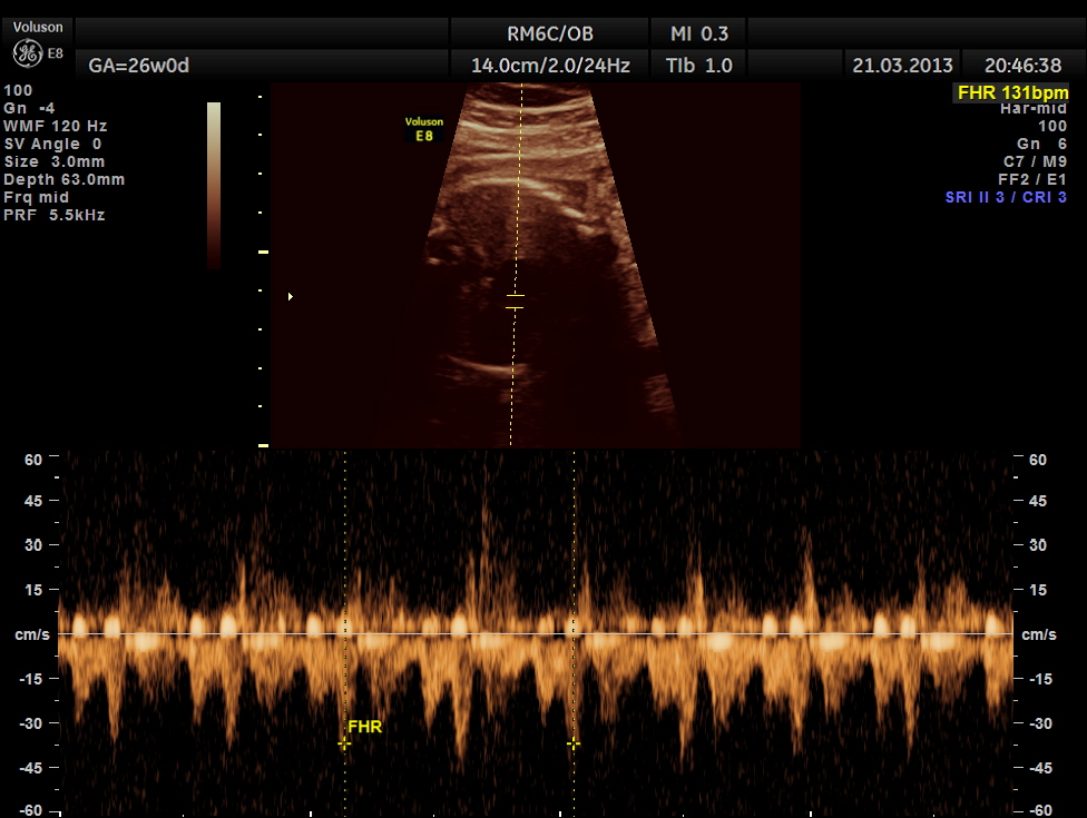
this shows a normal conduction and rate
A V VALVES are at the same level
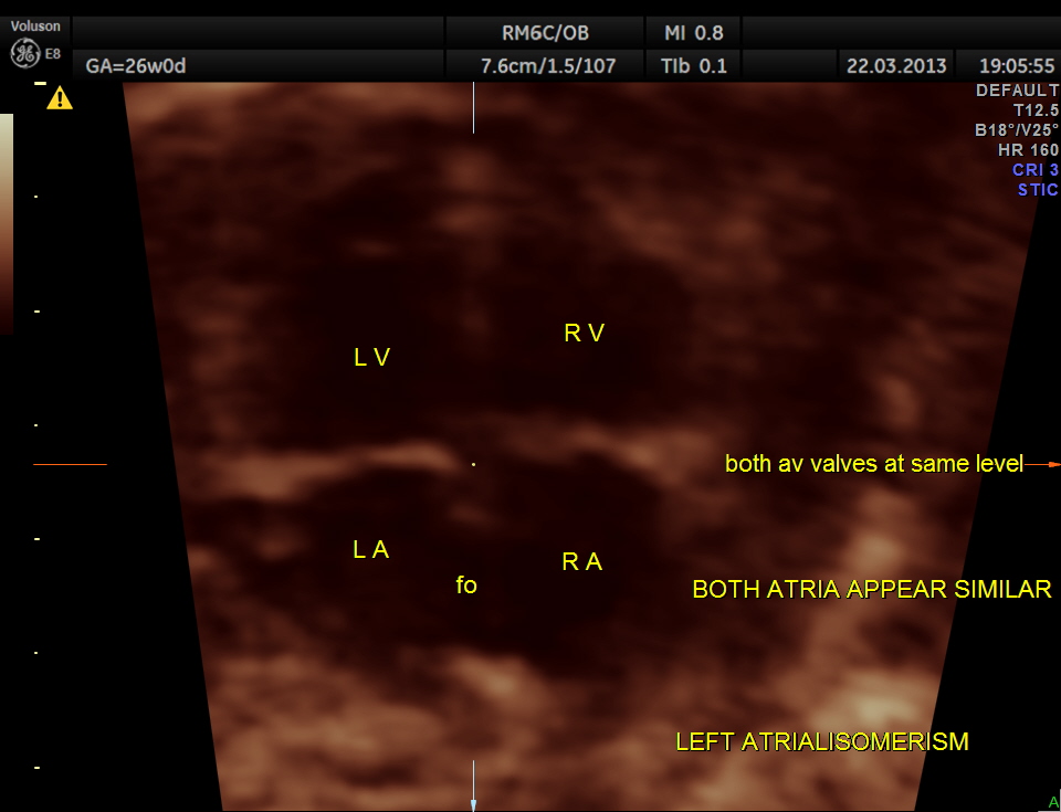
A V VALVES AT SAME LEVEL
STIC image shows ATRIO VENTRICULAR SEPTAL DEFECT ( ENDOCARDIAL CUSHION DEFECT )
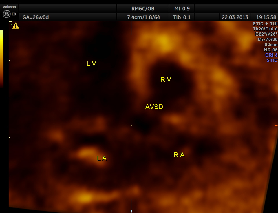
STIC image showing A V SEPTAL DEFECT
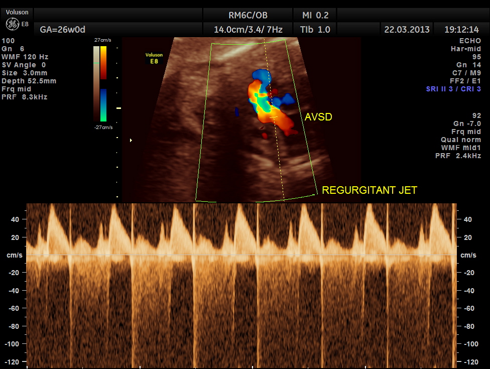
The next image shows spectral doppler showing prominent regurgitant jet in A V SEPTAL DEFECT
To summarise this fetus had
1. double vessel sign of aorta and azygos vein running side by side , with azygos slightly posterior,
2. irregular rhythm of A.V.Dissociation, Bradycardia and normal rhythm
3.Atrio ventricular septal defect ( endocardial cushion defect )
4. Prominent regurgitant jet seen at A V septal defect
5. Abnormal cardiac axis
This was referred for detailed evaluation as the irregular rhythm was picked up at a different centre.
Very useful links are given below.
The Fetus.Net : Left atrial isomerism
Dedicated to the mission of bringing free or low-cost educational materials and information to the global ultrasound community

Pingback: LEFT ATRIAL ISOMERISM , SITUS AMBIGUUS (or AMBIGUOUS), HETEROTAXY | When I turned 53
Pingback: TETRALOGY OF FALLOT WITH RENAL DYSPLASIA ( FOLLOW UP AFTER BIRTH ) | Looking Through a Transducer