This was a 25 year old lady ,with history of consanguinity was gravida 4, para 2, miscarriage 1 , live 1 ; Her 1st child died at 7 months of age and was diagnosed to have non obstructive hypertrophic cardiomyopathy causing cardiac failure. The hospital had suspected mucopolysacharidoses as a possibility . Her 2nd child is supposedly normal.
She was referred for evaluation of the fetal heart.
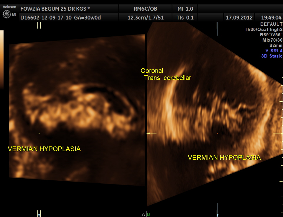
Ultrasound pictures of the fetal cranium are given below and showed cerebellar vermian dysgenesis .The buttocks sign can be appreciated.

![]()


3 D RECONSTRUCTION
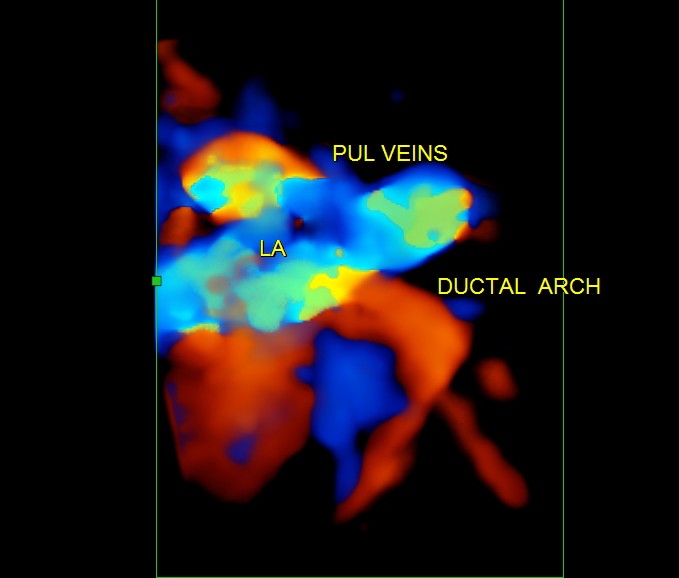
The next 2 images show the findings of the fetal heart. Significant hypertrophy of the septal and apical regions of the Left ventricle are seen.


This patient obviously needs further follow up and work up ; i will try to give the details later.

hi krish how about collecting all your foetal diagnostic images & coming out with a book? I think you should keep projecting in local Vernaculars not only to improve ur image but also to make others including our colleagues to know how imaging has improved in leaps & bounds…..;-)
LikeLike
Hi Deiveegan ,
Thanks for your comments
KRISHNAN
LikeLike