
A 45 year old lady with history of hysterectomy and bilateral salpingho oophorectomy, presented to her gynaecologist for symptoms of lower urinary tract – dysuria and frequency.
She was sent for a routine evaluation of the abdomen and pelvis.
The following pictures show a normal liver, gall bladder, pancreas and spleen .


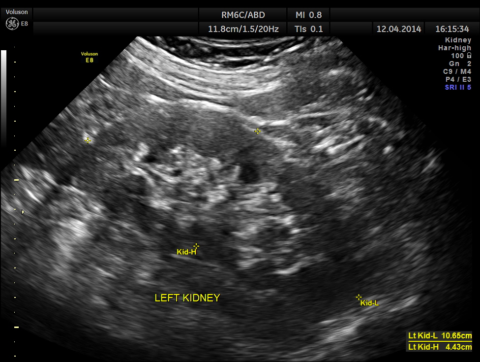
Both the kidneys appeared to be normal . No calculus was seen . There was no evidence of any obstruction.

Urinary bladder wall was mildly thickened . 3D showed fairly normal bladder mucosa . Ureteric jets were seen normally.
The distal ureters were not visualised .


Post void bladder was studied and showed the following .

Lo and behold – a distal right ureteric calculus is clearly seen now .

Usually we pick up all ureteric calculi and distal ureteric pathologies with a full bladder . Usually distal ureteric calculus will cause some amount of obstructive features in the ureter and the kidney . That was also absent in this patient .
But occasionally like this patient , the distal ureters can be compressed with a full bladder and such findings could be missed unless we do a post void study, especially when they have a LUTS symptoms . In this patient the bladder wall also showed mild thickening.

Many thanks for posting a case with academic interest.
One Question :
You have measured both pre & post void volume in a single scan . We usually take two scan one in Ts & other in Ls for each volume measurement. Which one is more reliable single or double scan ?
LikeLike
Definitely double ; you are right
LikeLike
Pingback: Lithiase urinaire : colique néphrétique simple, fébrile et hydronéphrose | thoracotomie
Interesting case, tempting to do a quick ultrasound and asks a complementary CT scan instead of doing a complete study in echography
LikeLike
When US does not give an answer , a complimentary CT is ideal ; but what I wanted to stress was the fact,that at times a post void bladder brings out the ureteric pathology . Thanks for your comments
LikeLike
One C.T.Lower Abdomen would have resolved the problem instead of taking too many pictures giving pain and expense to the old lady, am I right krish?
LikeLike
I always thought CT was costlier than USG. One has to consider radiation also. When US does not give an answer , a complimentary CT is ideal ; but what I wanted to stress was the fact,that at times a post void bladder brings out the ureteric pathology
LikeLike
Very interesting! Thanks for sharing this case. I will keep this in mind.
LikeLike
intresting
LikeLike
Calculi under 7 mm are so difficult to identify with ultrasound as hard to achieve posterior shadowing. Well done!
LikeLike
Thanks
LikeLike
Interesting case….thank you Kriznan!
LikeLike