
This was a 32-year-old primigravida who came for a routine anomaly scan around 22 weeks of gestation.
She had undergone 2 scans at 6 weeks and 8 weeks of gestation. A single fetus was reported .
Now she had the following findings.
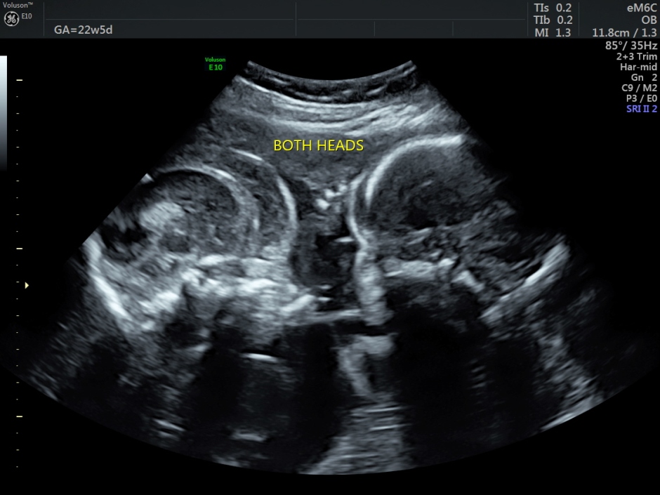
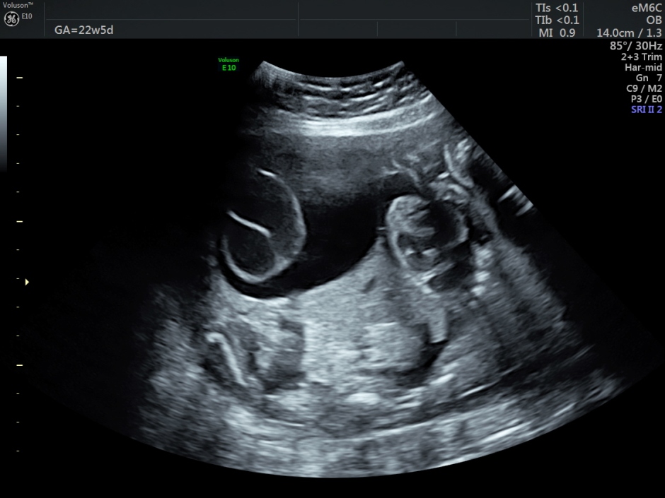
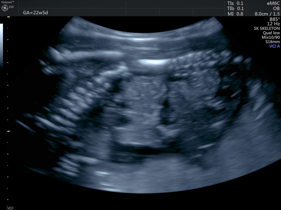
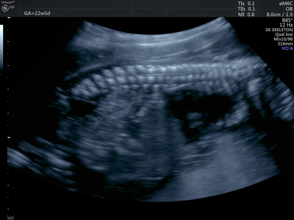
Two heads were seen.
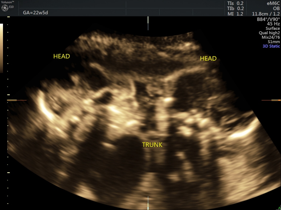
A single indistinct trunk was seen .
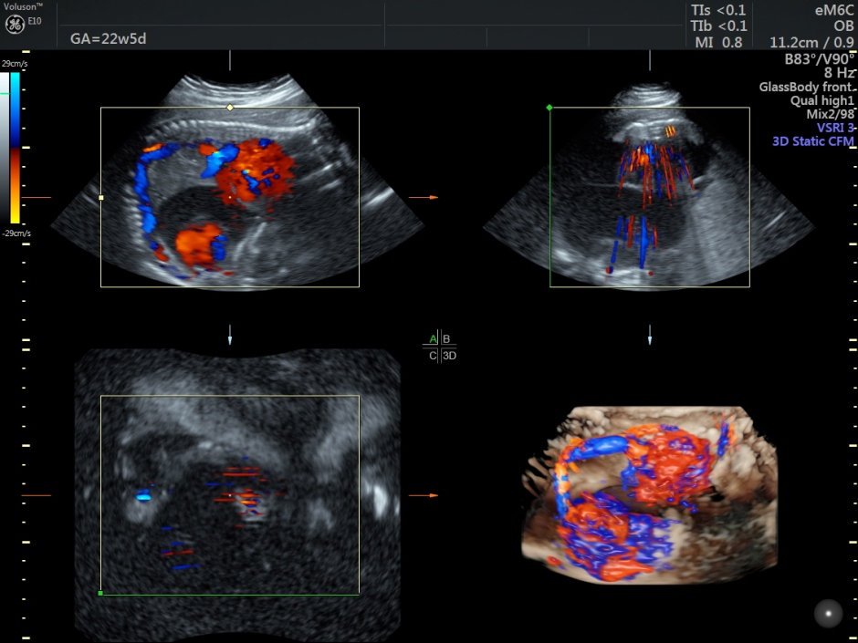
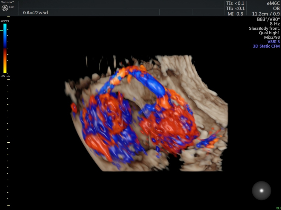
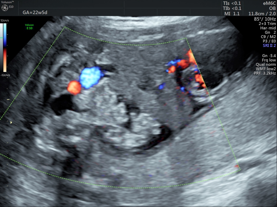
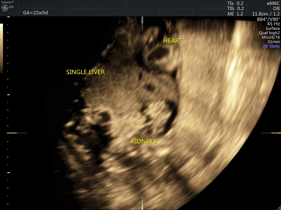
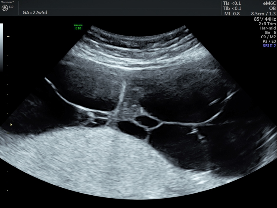
Two hearts were seen obliquely, inside a single chest.
Both showed severe pericardial effusion.
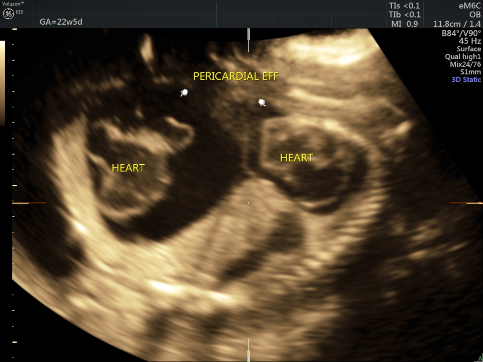
Both were probably connected by some vascular channel.
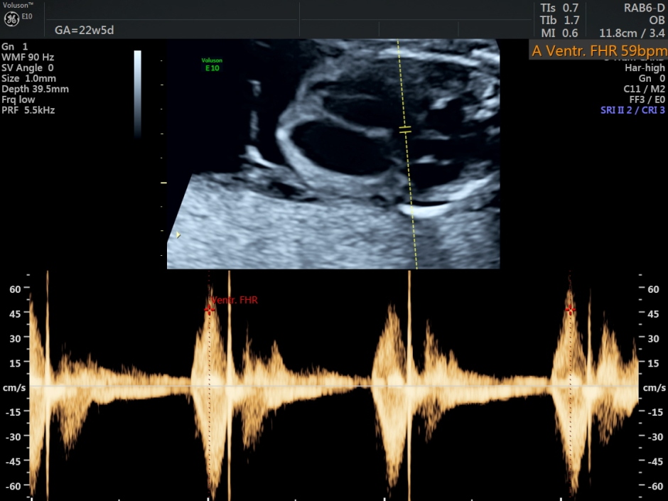
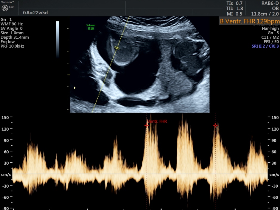
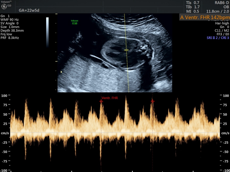
Fetal heart rate was variable from severe bradycardia to normal.
A single midline liver was seen.
Stomach bubble was not seen separately.
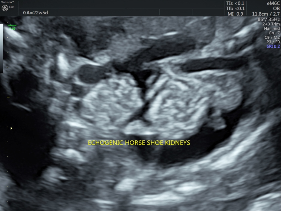
The fetal kidneys were brightly echogenic and showed a horseshoe appearance.
The urinary bladder was not seen clearly.
Multiple limb buds were seen.
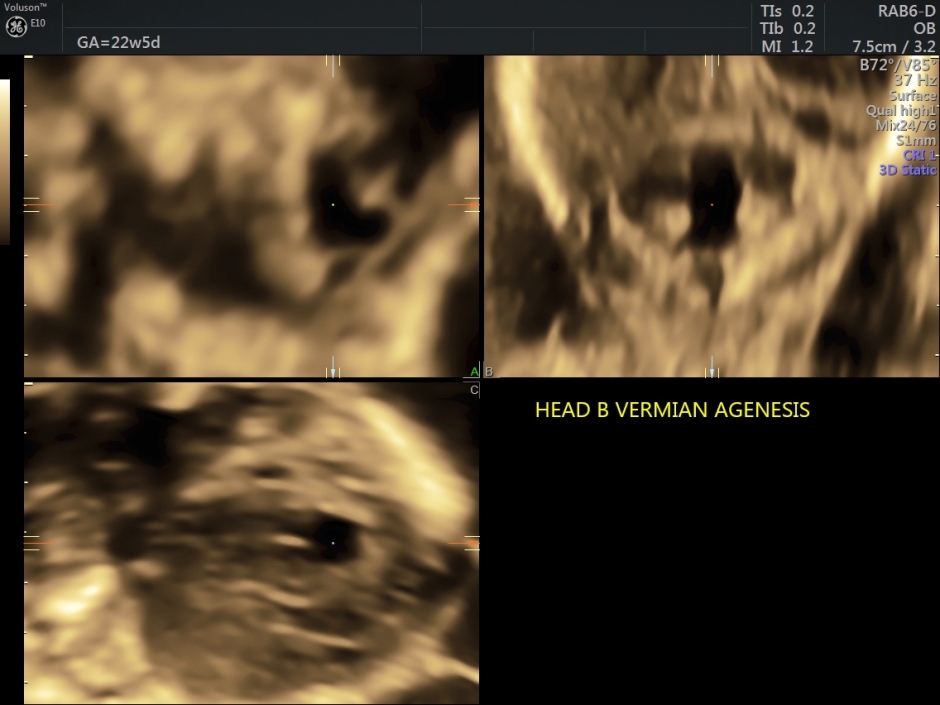
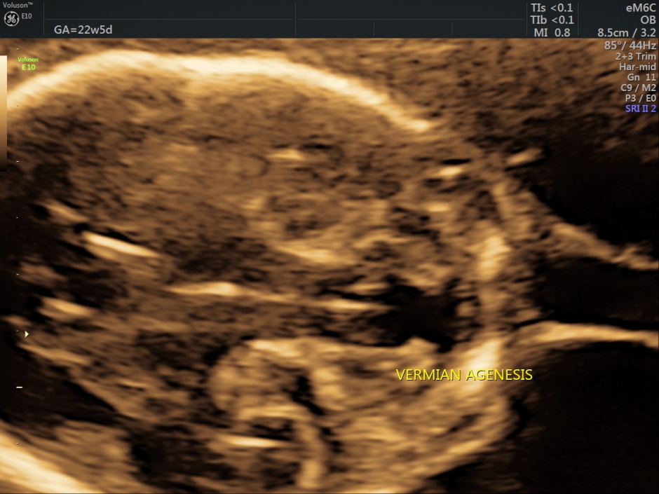
On more detailed organ evaluation, the head on the right showed vermian agenesis.
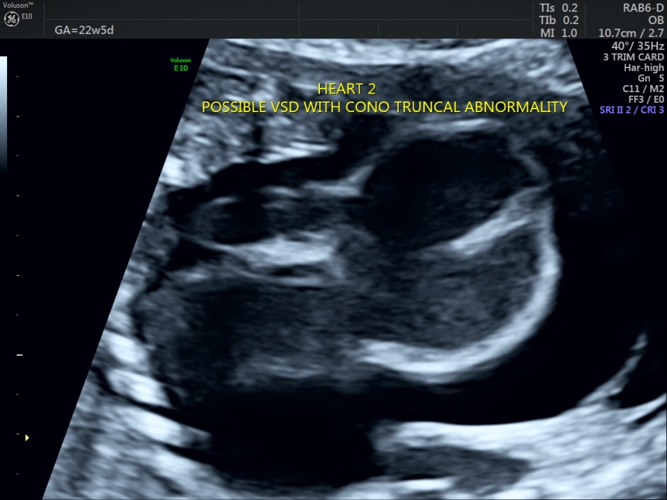
The heart on the right side showed possible VSD and a conotruncal abnormality, with a single large vessel arising from the heart.
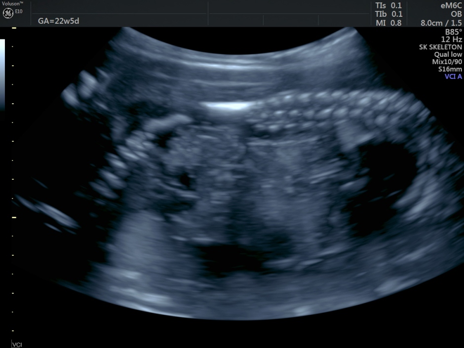
The fetus showed 2 spines connected at the lower end with an obtuse angle between them.
Amniotic fluid was reduced . Severe edema of the fetuses was seen. Multiple septations were seen in the fluid . – ? associated CYSTIC HYGROMA OF THE RIGHT SIDED FETUS.
The diagnosis offered was conjoined twins with a single trunk, two heads, and multiple limbs.
The fetus on the right side showed Vermian agenesis ,VSD with Conotruncal abnormality – probable truncus arteriosus and ? cystic hygroma.
Severe pericardial effusion was seen around both hearts.
Horseshoe kidneys were seen,
Stomach and Urinary bladder were not visualized.
The spine was connected at the lower end at an obtuse angle.
Limbs were seen, but difficult to ascertain number.
The following links would give more information.
http://umm.edu/programs/conjoined-twins/facts-about-the-twins
https://sonoworld.com/TheFetus/page.aspx?id=283
The following is taken from the above two web sites .
Conjoined twins are genetically identical, and are, therefore, always the same sex. They develop from the same fertilized egg, and they share the same amniotic cavity and placenta. MONOZYGOTIC, MONOAMNIOTIC, AND MONOCHORIONIC.
Twinning occurs one of two ways:
- either a woman releases two eggs instead of the usual one. If she releases two eggs, which are fertilized by separate sperm, she has fraternal twins.
- or she produces only one egg that divides after fertilization. When a single, fertilized egg divides and separates, she has identical or paternal twins.
In the case of conjoined twins, a woman only produces a single egg, which does not fully separate after fertilization. The developing embryo starts to split into identical twins during the first few weeks after conception but stops before the process is complete. The partially separated egg develops into a conjoined fetus.
Births of conjoined twins, whose skin and internal organs are fused together, are rare. Conjoined twins occur once every 200,000 live births, and their survival is anything but assured.
Approximately 40 to 60 percent of conjoined twins arrive stillborn, and about 35 percent survive only one day. The overall survival rate of conjoined twins is somewhere between 5 percent and 25 percent.
For some reason, female siblings seem to have a better shot at survival than their male counterparts. Although more male twins conjoin in the womb than female twins, females are three times as likely as males to be born alive. Approximately 70 percent of all conjoined twins are girls.
The following diagram is from fetus.net – from Phillippe Jeanty
Fig. Schematic drawing demonstrating the outcome of twinning at different stages of early embryonic life.
Top: Fission before the formation of the inner cell mass and any differentiation will produce two embryos with two separate chorions, amnions and placentas.
Middle: Twinning at the early blastocyst stage, after formation of the inner cell mass, will cause the development of two embryos, with one placenta and one chorion but two separate amnions.
Bottom: If separation occurs after the formation of the embryonic disc, the amnion has already formed, and will lead to a monoamniotic, monochorionic pregnancy. Incomplete fission at this stage or later will result in conjoined twins).
If, however, twinning is not initiated until after the embryonic disc is formed, only one amniotic and chorionic sac would develop, containing both twins. If the centers of embryonic growth do not separate adequately, it could be assumed that the transitional regions would be shared by the two embryos and would develop into conjoined twins.
A different explanation states that splitting of the original embryonic area occasionally takes place and that the extent and site of the splitting determine the different varieties of conjoined twins2. The existence of separate or conjoined twins is generally determined prior to the end of the second week following fertilization.

Thanks for the knowledge share Kriz
I am also seeing for the first time an USG image of twins.
Conjoined twins wow……
Great.
LikeLike
thanks for sharing and details krish.
LikeLike
Thanks Kriznan, great pictures (as always) showing the difficulties of trying to sort out the anatomy in later gestations! I have seen 8 sets of conjoined twins in my time, none of whom survived and ranging from 12 to 34 weeks gestation @ diagnosis. Your case illustrates the “missed opportunities” of very early scanning! Currently advised routine 12 week scanning would have picked this up.
LikeLike
Thanks
LikeLike
Very interesting case. Thank you for sharing Kriznan!
LikeLike
It’s too bad this was not discovered sooner for the patients sake. I am a sonograher and recently found conjoined twins at 9.5 weeks GA. Shared heart and chest down to unbilicus, cystic hygromas already able to be noted along with hydrops. This news would never be easy to give or get but the earlier the better I’d imagine.
LikeLike
I agree with you
LikeLike
Very interesting case, thanks for sharing.
LikeLike
Wonderful work keep sharing this will be useful to so many in this field
LikeLike
we found a conjoined twin in CNMU Rangpur,Bangladesh and published as case report in the ASEAN J of Radiology
LikeLike
Siamese twins were identified by a colleague at 10 weeks. 8 weeks should have been sufficient for screening of this pregnancy described above. But if there were two heartbeats seen and two supposed bodies seen, why wasn’t a 14/16 week ultrasound performed? Q4weeks per twins, typically and cervical length.
LikeLike
The 6 and 8 weeks scan were done elsewhere. Two heartbeats were not reported at that time. In all fairness to the doctor, the patient was advised a scan around 13 weeks. The patient ignored that and came for a scan now
LikeLike
Gotta love non compliance
LikeLike
In India non compliance is high
LikeLike
You have shown non compliance to be an issue in some of your other cases. Very frustrating for a perfectionist like yourself Kriznan!
LikeLike
Very interesting. I learned from this. I have
Never seen conjoined twins so artical was very
Helpful. Thanks for sharing
LikeLike