
Recently as I was hoping to wind up work after a long day, I received a call from my orthopaedician friend asking me to do an urgent scan.
This was a 36 year old man who fell down from a tractor and it ran over his left thigh . He was taken to an ortho consultant in a state of shock . After stabilisation , the left lower limb pulsations were not felt from the popliteal artery downwards.
He was sent for an emergency scan to assess the left lower limb arteries.
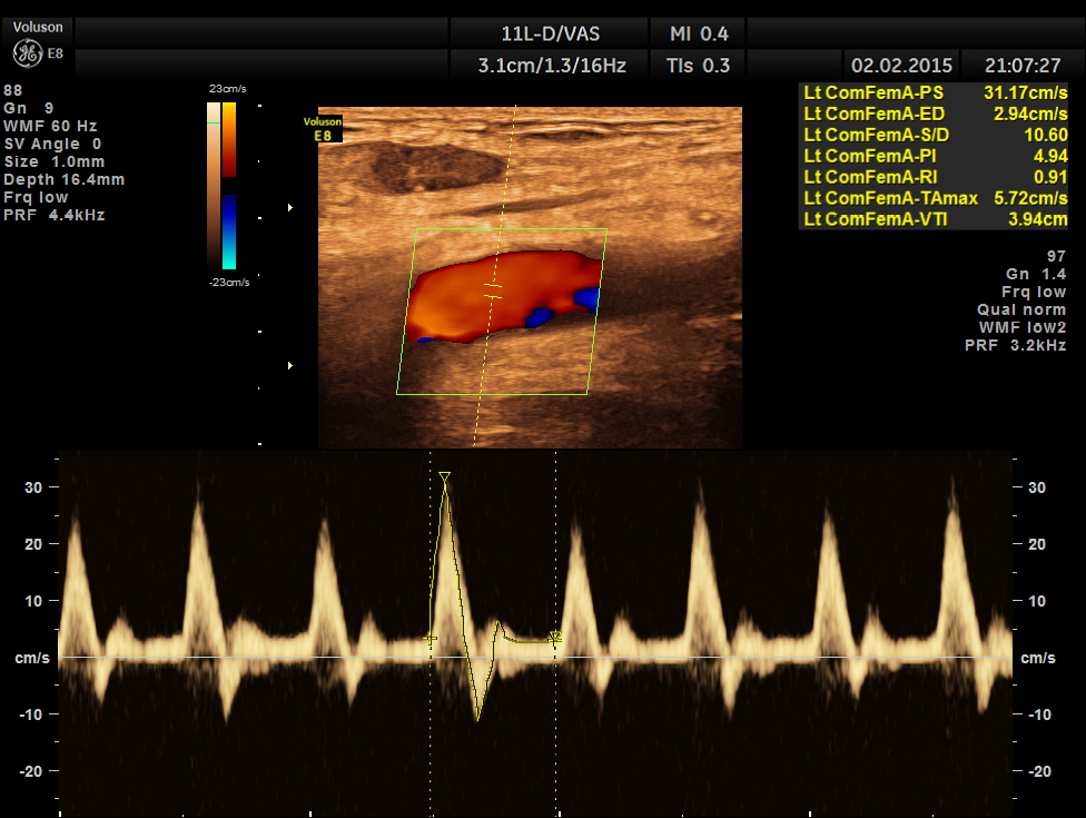
the left common femoral artery showed normal flow
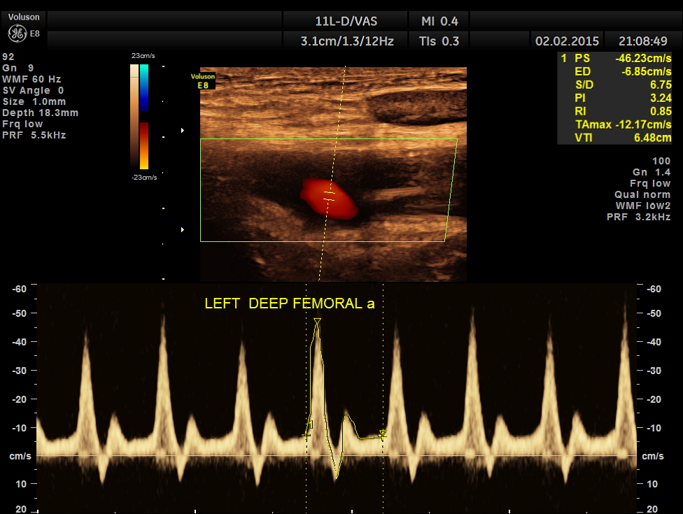
left profundal femoral artery shows normal flow
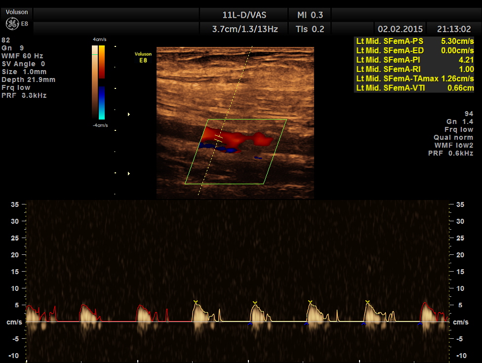
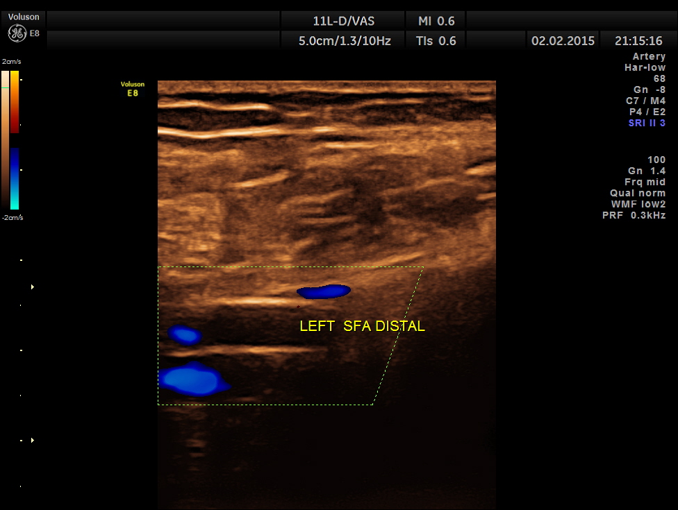
left proximal superficial femoral artery shows reduced peak systolic velocity
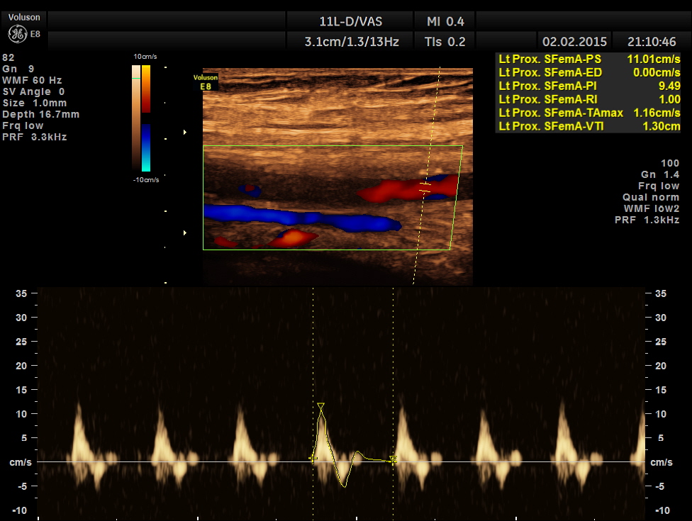
the following pictures show progressive fall of peak systolic velocity with no flow made out in the distal superficial femoral artery.

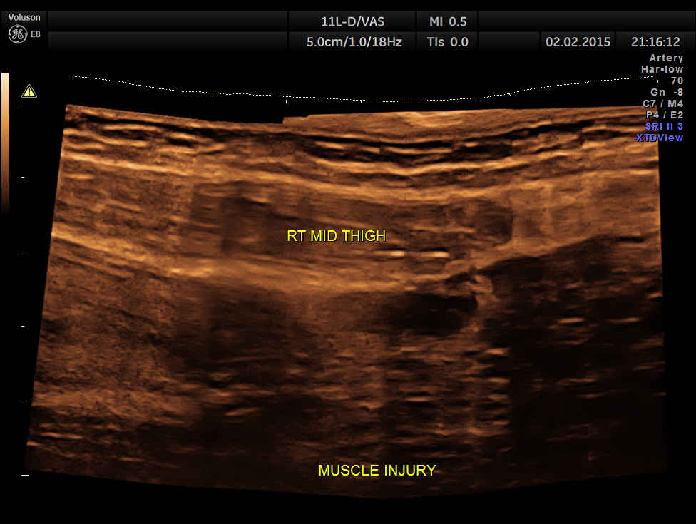
the severely damaged muscles of the thigh
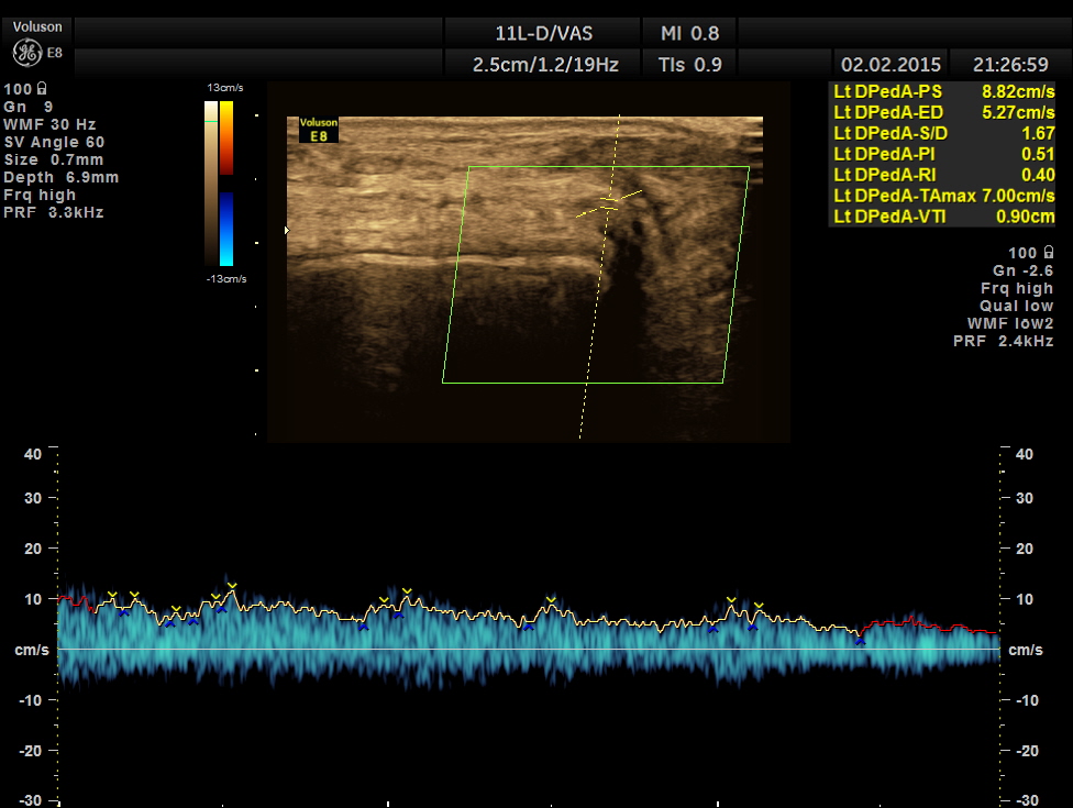
the popliteal fossa showed an artery with low flow monophasic flow, probably the popliteal artery

no flow was made out in the posterior tibial and dorsalis pedis arteries


i suggested that there could be an injury to the left superficial femoral artery , probably in the distal segment.
A vascular surgeon , who had already seen the patient felt exploration of that area was worthwhile and did that.
He found that there was a thrombus and a vessel tear beyond the adductor canal , close to the femoro popliteal junction . The thrombus was lysed with intra arterial heparin and the vessel was repaired . Within the next hour the patient regained pulsations in the distal arteries and in the next few hours the injured muscles also showed clinical improvement.
The next day my friend called me and said that his limb was salvaged.
The time frame is given below:
2.00 pm – the patient met with the accident
4.00 pm – he was seen by the orthopaedic specialist
8.00 pm – the surgical team wanted a vascular doppler assessment
9.30 pm – the doppler report was conveyed
1.30 am – the vascular surgeon started his work and completed in 3 hours time
I felt happy to have been part of this exciting work.

very nice kriz.. every time he feels the ground with his foot the credit goes to you & all those involved in limb salvaging work for that patient
LikeLike
Thanks Deiveegan
LikeLike
Wow, what an interesting case, glad that you and your staff were able to save the patient’s limb!
LikeLike
To clarify, all doctors involved are in different set ups ; glad we were able to help the patient
LikeLike
Intersting case Thank you for sharing this with the rest of us! Good team work!
LikeLike
Wow. Great case with a wonderful outcome. I enjoyed reading it.
LikeLike
Thanks
LikeLike
BRAVO, KRISHNAN Thanks,Elaine
LikeLike
you did an awesome vascular of study, you were instrumental in salvaging the patient’s limb! He didn’t develop a compartment syndrome.great case, great presentation. I’m curious as to why you don’t have 60 degree angle
l
LikeLike
Hello–thank you for posting this interesting case. I am assuming that you used no angle correction because it was an urgent exam? You certainly can see the waveform diminish in the SFA as you move inferior, however; the PW Doppler velocity measurements for the SFA are all erroneous. This must have been a difficult scan due to the patient’s pain? Thank you again for posting…
LikeLike
Thanks for your keen observations. I’m Happy to say the ultimate outcome was favourable to the patient. But I would agree with your observations. Thanks again
LikeLike
good
LikeLike