
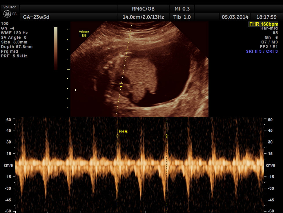
- This was a 22 year old lady, primi gravida who was referred for 2nd opinion for suspected fetal ascites.
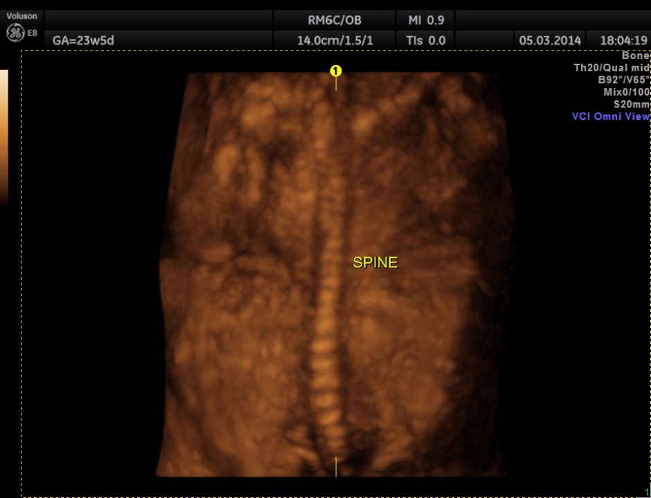
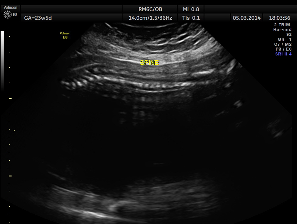
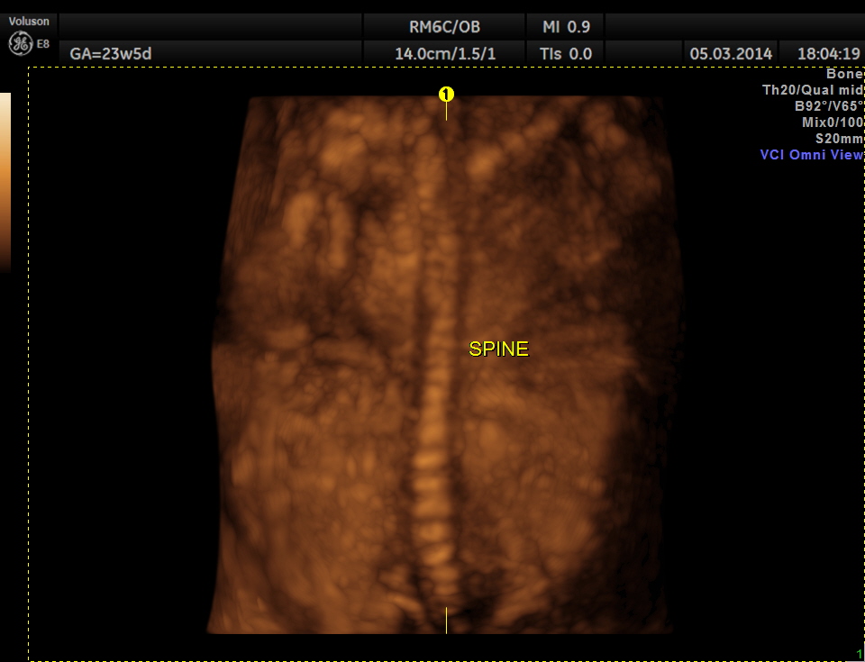
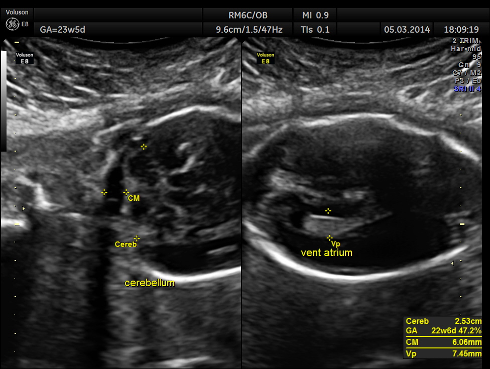
- The fetus was seen in a fixed position with spine fixed to the anterior abdominal wall. Kypho scoliosis could be made out.

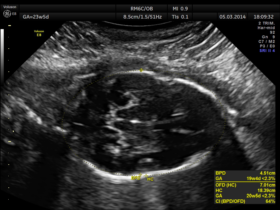
The head was in the upper pole and appeared dolichocephalic.
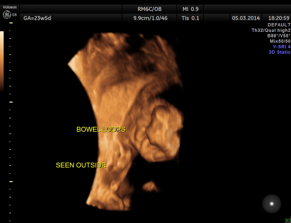
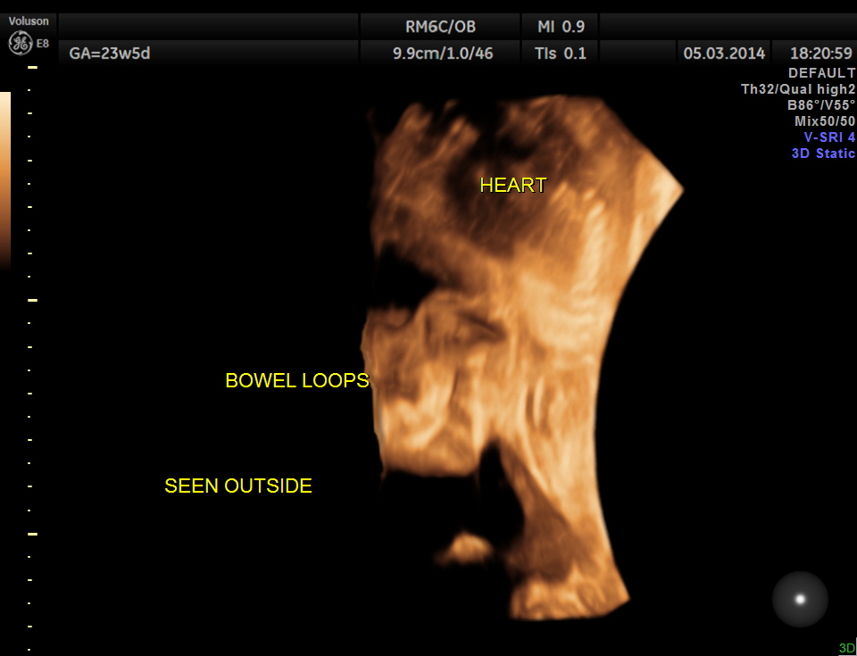
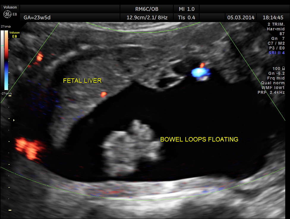
A large anterior abdominal wall defect was seen with the liver , bowels and the viscera floating outside .
Amniotic band is seen .

The following are a few 3 D reconstruction.
T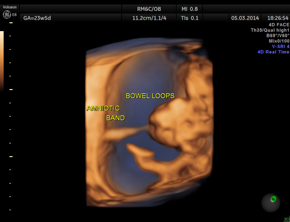

The following picture shows the reconstruction of the amniotic band.
t
The chest wall seems to be intact in the pictures below.

The fetal kidneys with the renal arteries are shown in the picture below.

The abdominal wall defect was extensive and the lower abdominal wall is also not made out.
? Cloacal exstrophy is seen below.

Floating viscera are seen .


The diagnosis I offered on that day was anterior abdominal wall defect – body stalk complex with amniotic band with kyphoscoliosis of the spine.
Abdominal wall defects http://radiopaedia.org/articles/fetal-anterior-abdominal-wall-defects
Fetal anterior abdominal wall defects can occur with a number of pathologies and include
Individual entities
Syndromes / complexes
- limb body wall complex
- OEIS complex
- omphalocoele-radial ray (ORR) complex
- Pentalogy of Cantrell
- amniotic band syndrome : if there are band through the anterior abdominal wall
Please visit some of the links given below
http://sonoworld.com/fetus/page.aspx?id=2420
Click to access IJRMS20130519.pdf
https://www.sonoworld.com/Fetus/page.aspx?id=2907
http://onlinelibrary.wiley.com/doi/10.1002/uog.84/pdf
The limb body wall complex (LBWC) is a rare variable group of congenital limb and body wall defects (involving mainly the chest and abdomen). They can include
Abdominal wall defect – abdominoschisis : usually large and left-sided 4 : almost always present
thoracic wall defect / thoracoschisis
ectopia cordis
anomalies of the lower limbs
clubfoot
brachydactyly
polydactyly
syndactyly
oligodactyly
absent limbs
scoliosis : often profound
exencephaly
Limb body wall complex is a rare syndrome with two distinct phenotypes described by Russo et al. in 1993: a form with a placento-cephalic attachment and another form with a placento-abdominal attachment. Diagnosis is based on various echographic signs. No case of postnatal survival is described.
Pathogenesis of LBWC is unclear and uncertain. Three pathogenic theories have been proposed:
1. Early amnion rupture theory (Exogenic theory) leading to the formation of amniotic bands that interrupt embryogenesis and the fetus lies outside the amniotic
cavity. It disturbs the normal development (Torpin et al, 1965).
2. Vascular disruption theory (Endogenous theory) is described as events that negatively influence normal embryonic blood supply, thereby interrupting normal
morphogenesis (Van Allan, 1987).
3. Embryonic dysgenesis or Germ disc defect with early embryonic maldevelopment. The fetal disruptions resulted from imperfect histogenesis, showing
disturbances of the embryonic folding process (Streeter theory 1930).
Body-stalk deformity occurs from complete failure of body folding along all three axes (cephalic, caudal, and lateral) . Normally after the embryo folds, the intraembryonic coelom (future peritoneal cavity) is separated from the extraembryonic coelom, and the umbilical cord subsequently forms . If this folding does not occur, then the extraembryonic cavity is not obliterated and the body-stalk is missing. This leads to congenital absence of the umbilical cord with a wide-based, peripheral insertion of the large fetal amnioperitoneal sac onto the placental chorionic plate . Aberrant cephalic folding can further produce ectopia cordis congenital heart disease, diaphragmatic hernia with resultant pulmonary hypoplasia, and a defect in the thoracic wall and epigastrium. The lack of lateral folding is the basis for herniation of the midabdominal contents into the amnioperitoneal sac, while abnormal caudal folding results in the malformations noted with cloacal exstrophy. Severe scoliosis develops as a consequence of the irregular attachment of the fetus to the placenta
Other possible mechanisms for body-stalk malformation include: decreased blood flow early in gestation (four to six weeks) leading to disruption and incomplete development of embryonic tissue and early amnion rupture (before obliteration of the coelomic cavity) allowing the fetus to pass into the coelomic cavity . In the latter, direct mechanical pressure and amniotic band formation are thought to create the associated defects.
Body-stalk anomalies are the rarest of all abdominal wall defects. The average incidence is 1 in 14,273 births

kudos to you Kriz. most of what you show is greek & latin to me. the explanation part nice. I think you should be getting ready to publish all this in a Book titled Ultrasonic findings of foetal abnormalities in India- from a clinic in a semi urban locality.
LikeLike
Hi Deiveegan ,thanks ; I am definitely planning to do that
LikeLike
thanks for reporting the rarest intrauterine developmental anamoly.
LikeLike
Amazing pictures Krishnan, and quite difficult to interpret from our seats! Good thing you explain things so well….
I remember my first body stalk complex in 1982… the machine had a monitor with only 64 lines. Wish I still had the pictures to show you the difference!
LikeLike
Thanks Patricia ; I ll have to admit that I look forward to your insightful comments
LikeLike
Thanks It mas very interesting!
LikeLike
Hi
Great case again! Haven’t seen one ( or similar in a long time.) Great images and reviews.
Thanks again, we need to see more than maybe we do in our own scope of things!
LikeLike
Quite helpful in medical education.
LikeLike
I enjoy your presentations and the images are excellent. Thank you.
LikeLike
Thank you
LikeLike
I found your entry while researching LBWC. I am the parent of a child who died shortly after birth, due to LBWC. We have 50+ parents in a group, who continued their pregnancies after this diagnosis- we generally have at least one parent joining us every two months. We are desperate to find if there is any research currently being done on this disorder. We would like to participate.
LikeLike
Sarah, I was about to post the same thing. Did you ever receive a response?
Jill
LikeLike
Hello Sarah &Jill,
I’m very sorry for not having responded long back. I’m not aware of the research going into the LBWC . If I know of anything I’ll let you know.
LikeLike
Thank you so much!
We did find researchers in the Netherlands (http://www.ncbi.nlm.nih.gov/pubmed/21472892) who are studying the PORCN gene, and who were willing to accept tissue samples from our parents, unfortunately the type of tissue they require is unable to be shipped internationally.
I am leaving my email here, in case anyone else finds this message thread.
info@limbbodywallcomplex.net
(we now have over 60 families… and members requesting admission regularly)
LikeLike