
This was a 58 year old man who saw a surgeon for complaints of dragging pain of the left scrotum.The surgeon’s clinical diagnosis was varicocele and he was sent for confirmation.
The following pictures show the lesion.
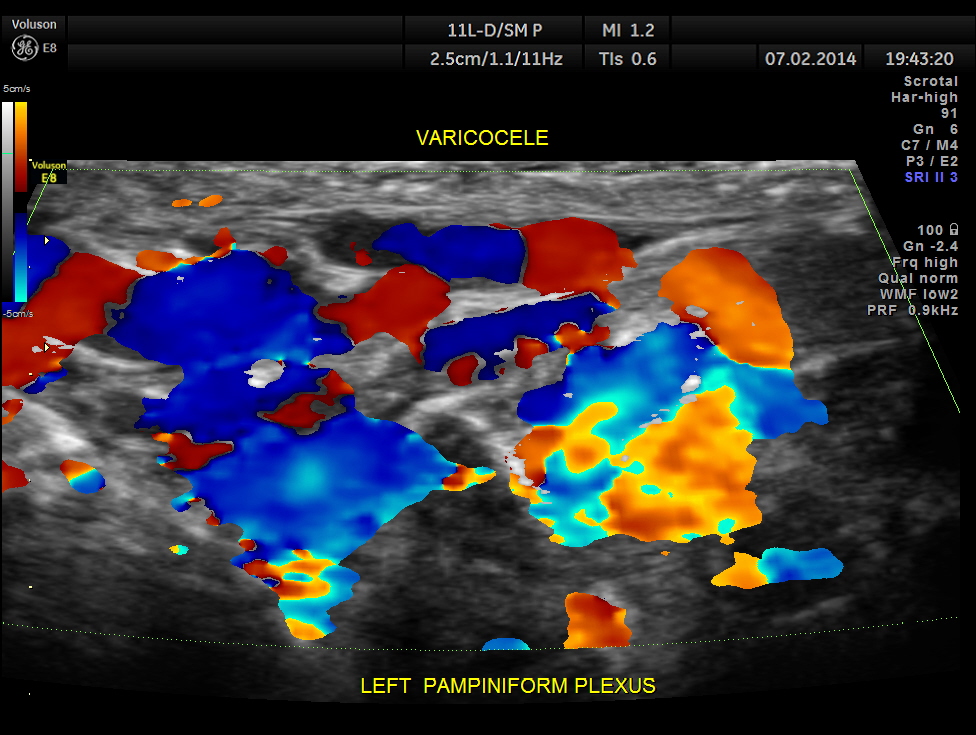
Prominent dilated veins seen in the left scrotum

COLOUR DOPPLER PICTURE GIVEN BELOW.
SPECTRAL DOPPLER SHOWN BELOW .
NORMAL RESPIRATION CAUSES FLOW REVERSAL.
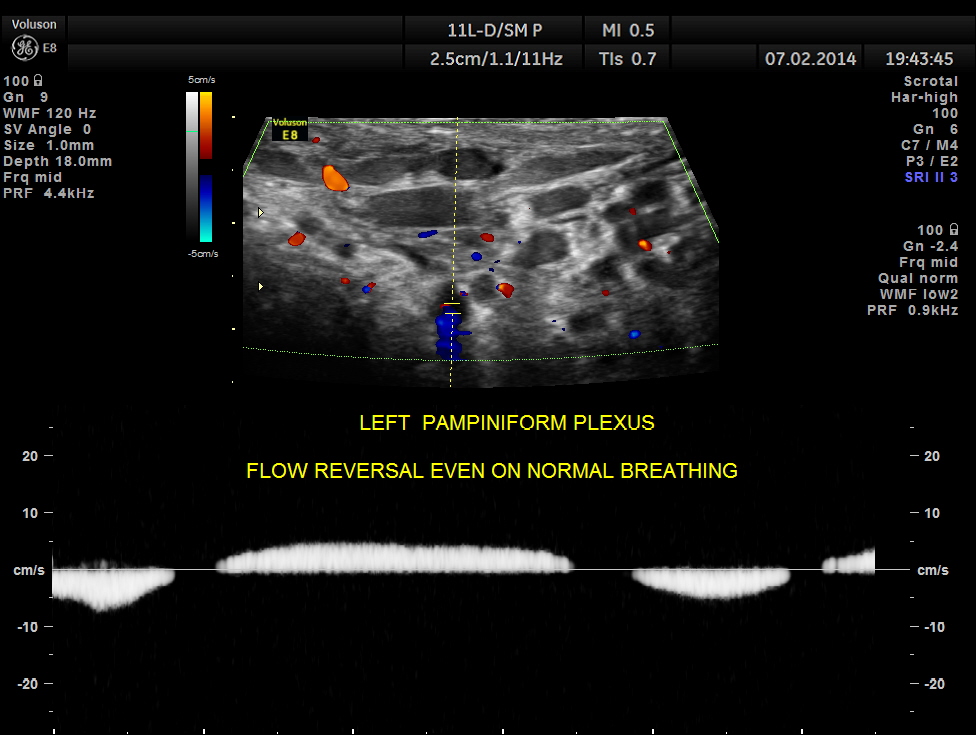
VALSALVA causes an exaggerated response.

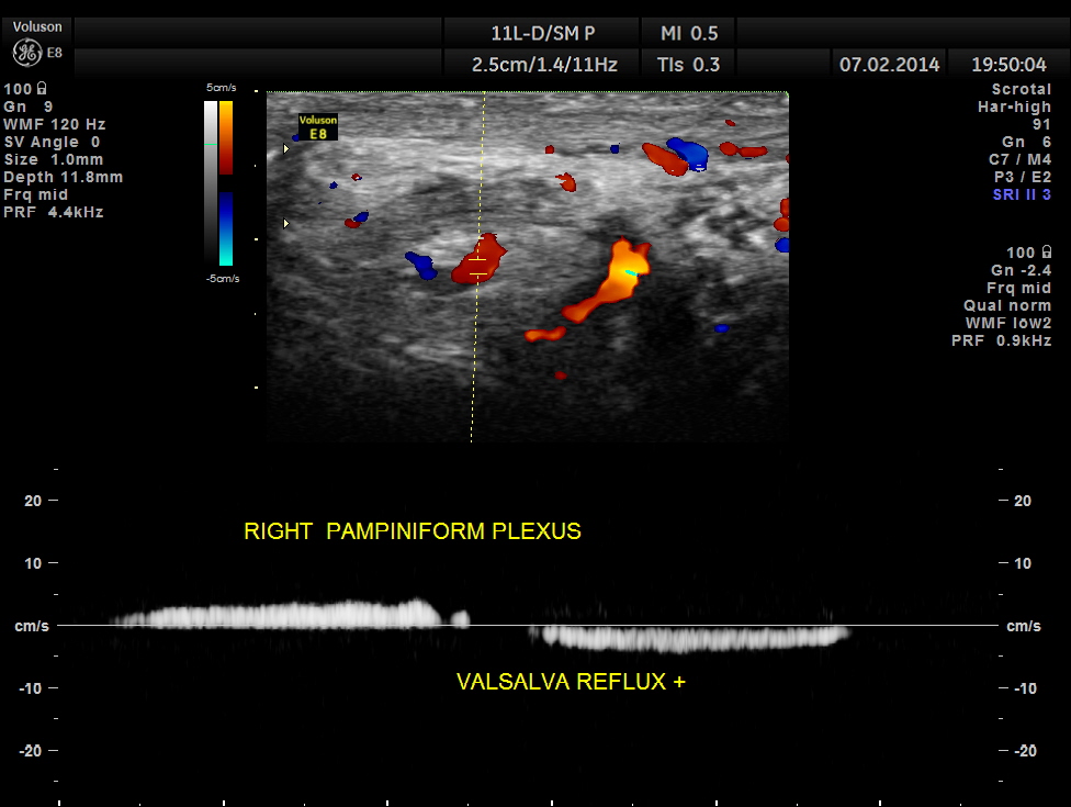
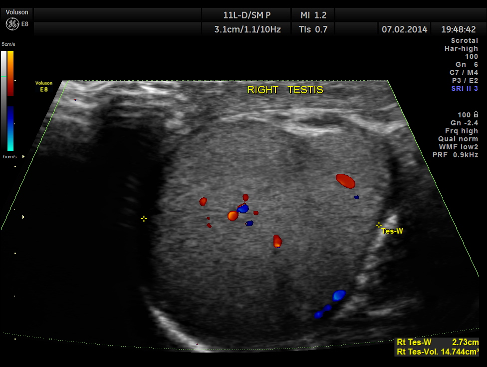
Right pampiniform plexus also showed dilated veins with reflux.

Spectral doppler shows reversal of flow .
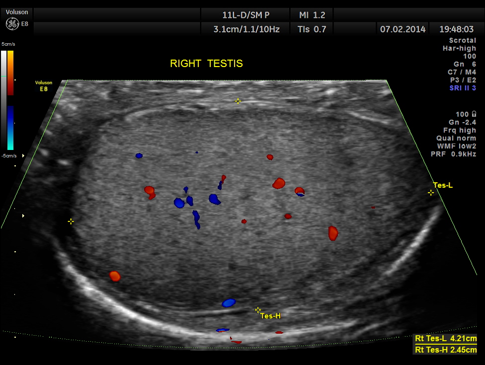
The following pictures show the testicles.Mild left hydrocele is also seen.


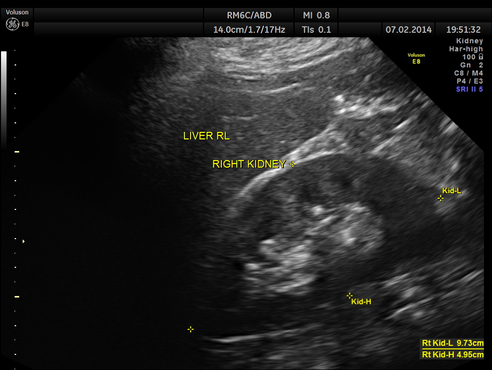
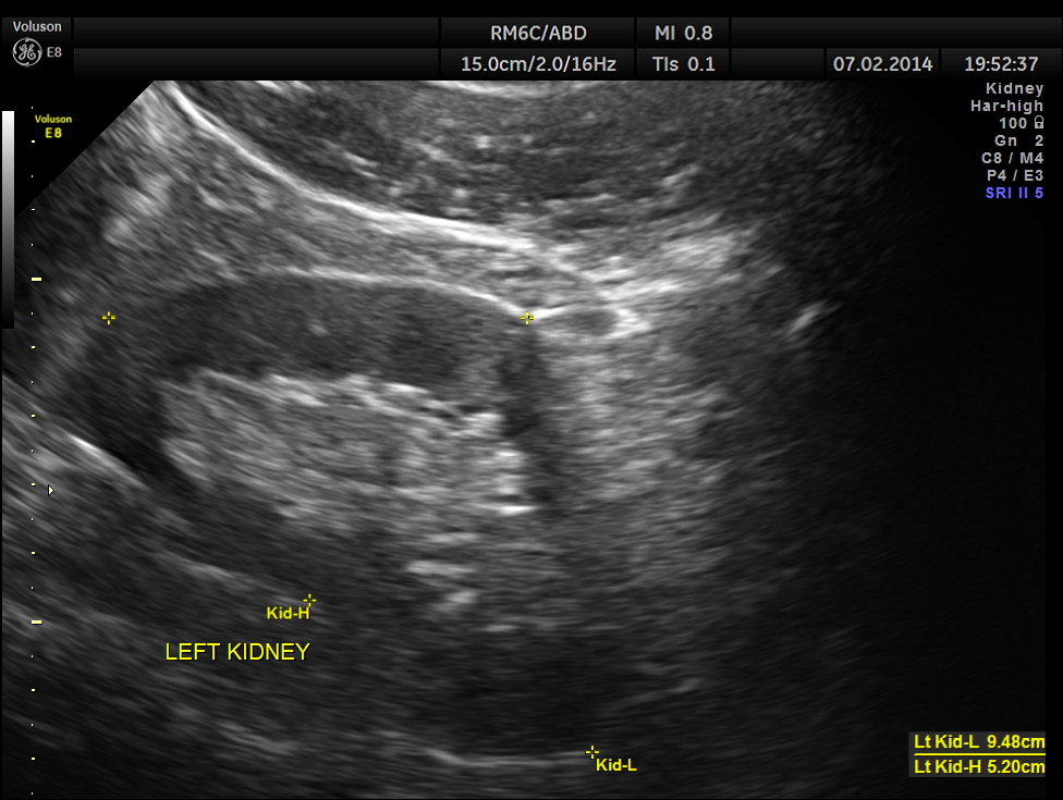
The kidneys appeared normal.
This patient had bilateral varicocele , left side being more prominent. Mild left hydrocele was also present.
The following link will open the wiki link.
http://en.wikipedia.org/wiki/Varicocele
Some interesting highlights from this are given below.
Varicocele is an abnormal enlargement of the pampiniform venous plexus in the scrotum. This plexus of veins drains the testicles. The testicular blood vessels originate in the abdomen and course down through the inguinal canal as part of thespermatic cord on their way to the testis. Upward flow of blood in the veins is ensured by small one-way valves that prevent backflow. Defective valves, or compression of the vein by a nearby structure, can cause dilatation of the testicular veins near the testis, leading to the formation of a varicocele.
Signs and symptoms
Symptoms of a varicocele may include:
- Dragging-like or aching pain within scrotum.
- Feeling of heaviness in the testicle(s)
- Atrophy (shrinking) of the testicle(s)
- Alteration of testosterone levels.
- Visible or palpable (able to be felt) enlarged vein
Cause
The idiopathic varicocele occurs when the valves within the veins along the spermatic cord do not work properly. This is essentially the same process as varicose veins, which are common in the legs. This results in backflow of blood into the pampiniform plexus and causes increased pressures, which on rare occasion can lead to permanent damage to the testicular tissue due to disruption of normal supply of oxygenated blood via the testicular artery.
Varicoceles develop slowly and may not have any symptoms. They are most frequently diagnosed when a patient is 15–30 years of age, and rarely develop after the age of 40. They occur in 15-20% of all males.
98% of idiopathic varicoceles occur on the left side, apparently because the left testicular vein connects to the renal vein (and does so at a 90-degree angle), while the right testicular vein drains at less than 90-degrees directly into the significantly larger inferior vena cava. Isolated right sided varicoceles are rare.
A secondary varicocele is due to compression of the venous drainage of the testicle. A pelvic or abdominal malignancy is a definite concern when a right-sided varicocele is newly diagnosed in a patient older than 40 years of age. One non-malignant cause of a secondary varicocele is the so-called “Nutcracker syndrome“, a condition in which the superior mesenteric artery compresses the left renal vein, causing increased pressures there to be transmitted retrograde into the left pampiniform plexus. The most common cause is renal cell carcinoma (a.k.a. hypernephroma) followed by retroperitoneal fibrosis or adhesions.

As usual :), very nice images Krishnan!
LikeLike
Thanks Patricia
LikeLike
good
LikeLike
a question on varicoceles: what is considered normal size (AP diameter) for pampiniform plexus veins? And is this during resting or straining state? I have the understanding it’s resting. I could be wrong.
LikeLike
Less than 2 mms would be normal ; > 3 mms will be dilated even in resting ; but will show an increase on valsalva
LikeLike
Thank you kriznan for those juicy images. I have never seen one before.also thanks for the exposition. Keep up the good work. Albert Nkemchor.
LikeLike
If a unilateral varicocele is seen, is it standard protocol to look at the kidneys? Or do you look at them anytime you see a bilateral or unilateral varicocele?
LikeLike
In anyone above ‘middle age’ I feel a look at the kidneys will be mandatory.
Unilateral varicocele , definitely yes
LikeLike