This was a 50-year-old gentleman , who presented with recurrent lower abdominal pain to the surgeon and was referred for a scan.
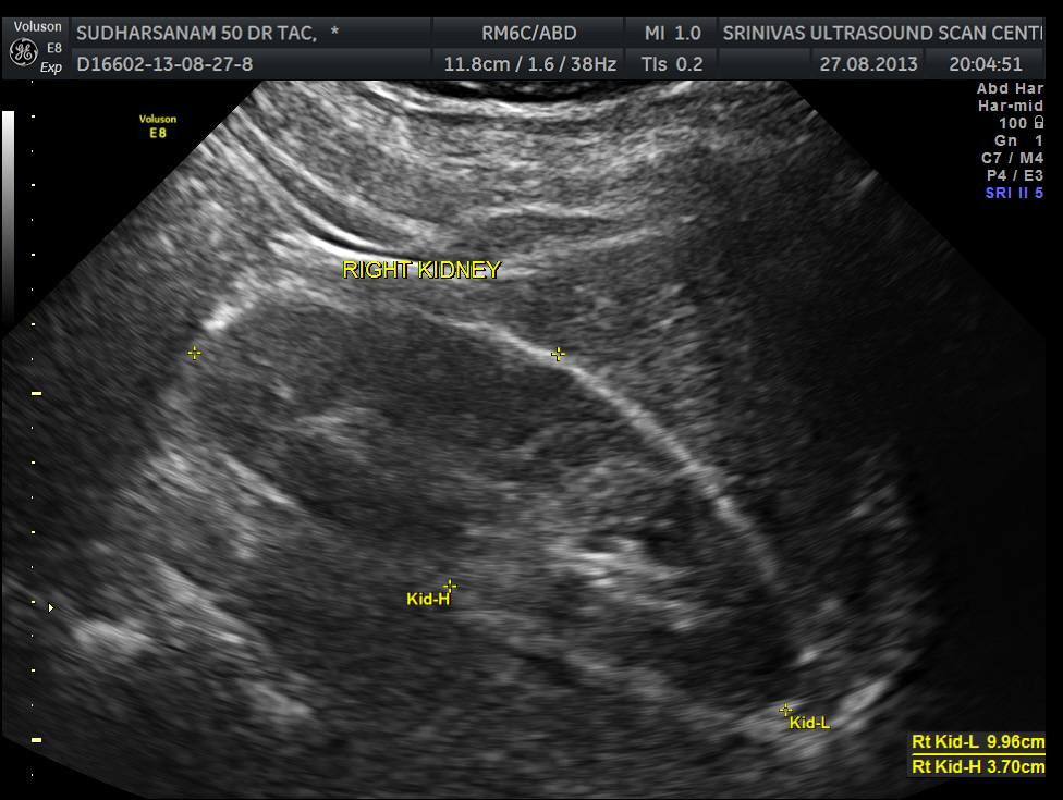
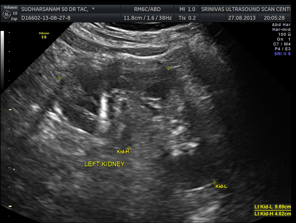
He had an essentially normal appearance of the upper abdominal organs . His kidneys also appeared to be normal.
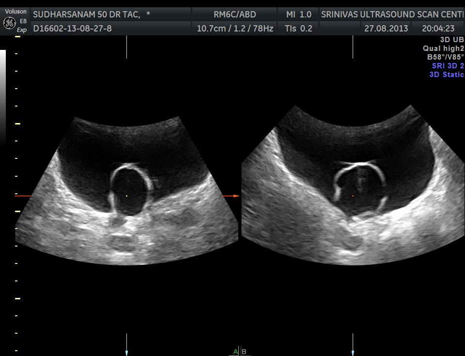
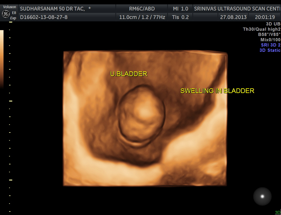
The urinary bladder showed the following :

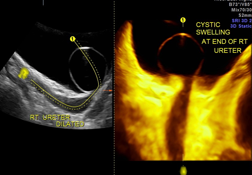
the picture on the left shows the dilated distal right ureter
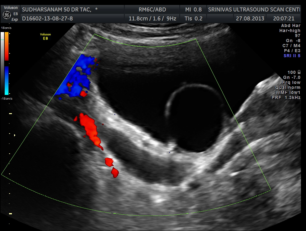
the dilated distal ureter is seen .Colour doppler shows flow in the adjacent vessels
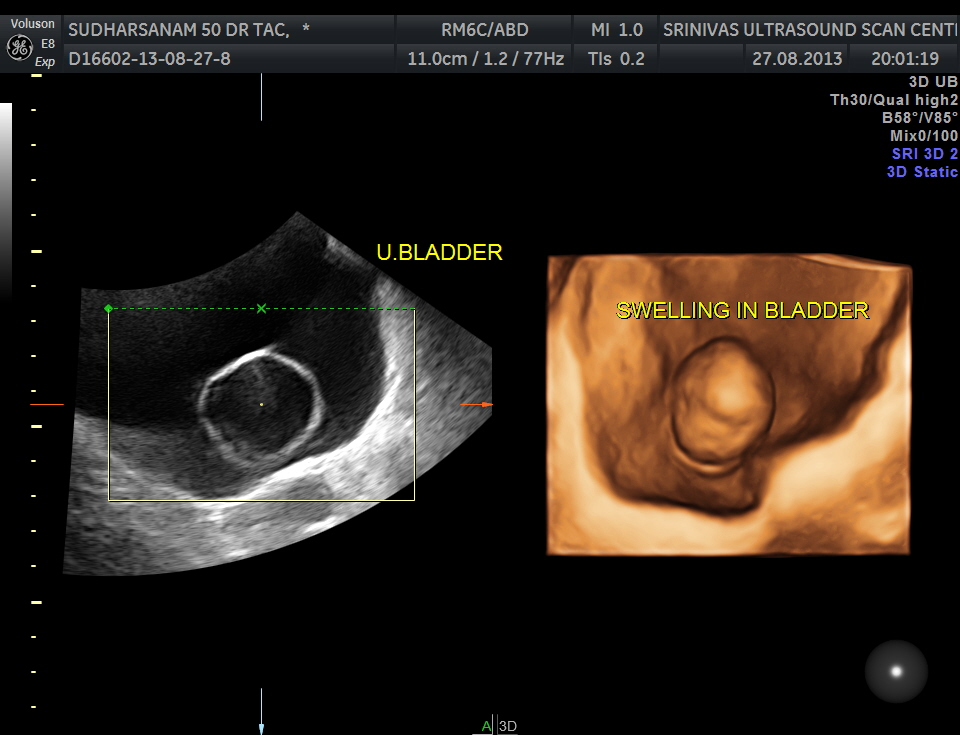
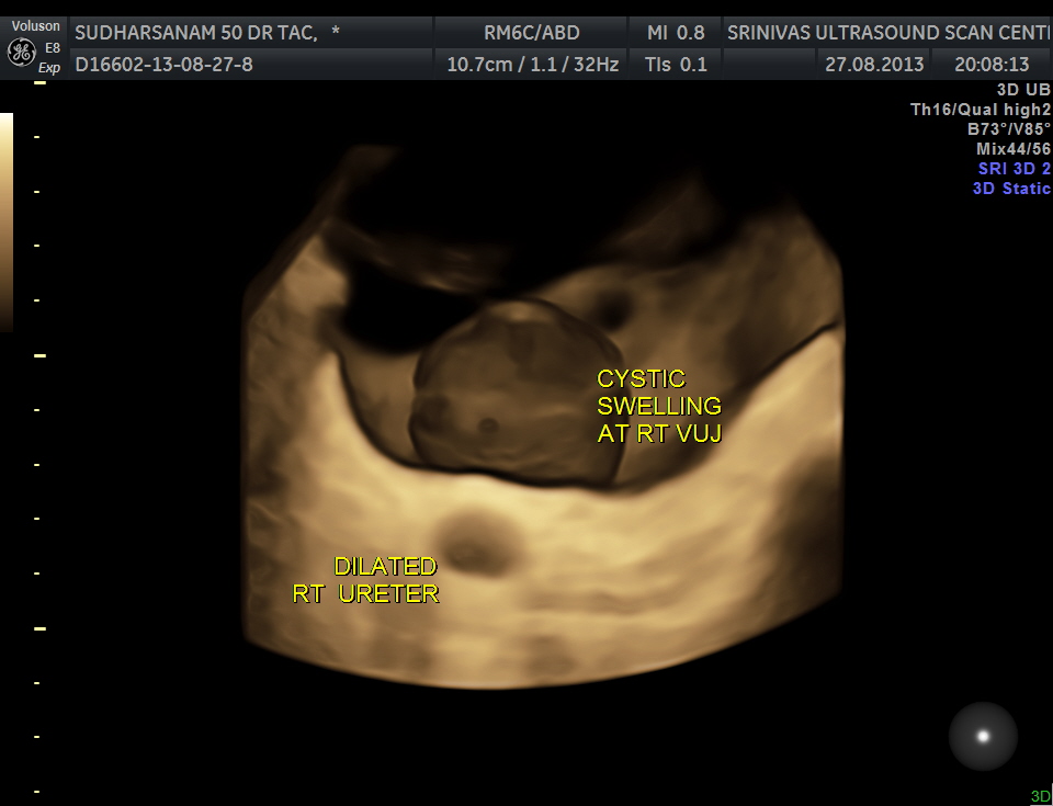
reconstructed image shows the swelling within the bladder
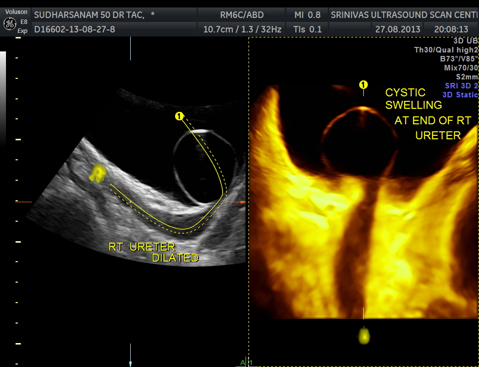
omni view shows the dilated ureter leading to the cystic swelling – Ureterocele
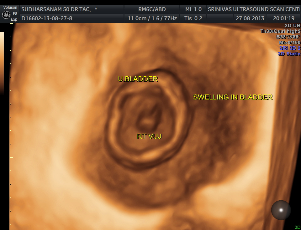
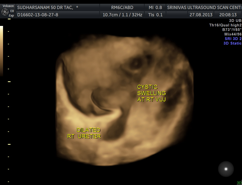
given below are few other reconstructions
images of the kidney are given below.
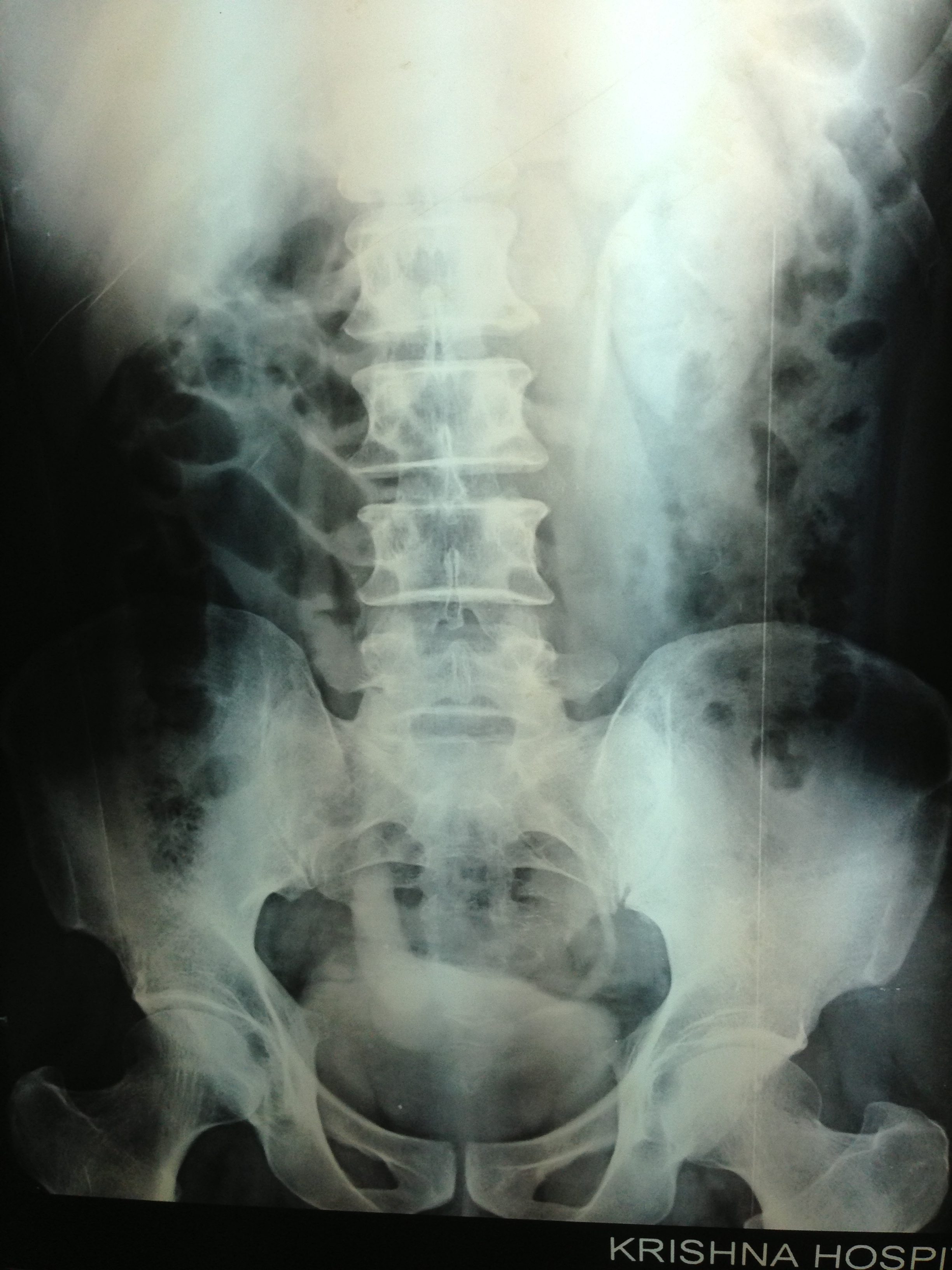
The patient was seen by the urologist and an IVP was done , which proved the diagnosis of orthotopic ureterocele . (a ureterocele entirely within the bladder.)
He has been advised cystoscopic surgical correction.

Excellent slides
LikeLike
Thanks sir
LikeLike
I have found many cases of ureterocele but not in 3D
LikeLike
Nice picks
LikeLike
Great complete case, images are outstanding.
Mike Keith
LikeLike
The images are very clear, thanks
LikeLike
Good images,well done!
LikeLike
In cases I have met the cyst disappears or reduces in size after voiding or passing urine. This has assisted me to confirm the diagnosis of a ureterocele
LikeLike
Thanks for sharing your experience
LikeLike
Dr Krishnan, once again an outstanding presentation of a case study with excellent pictures.Thanks for sharing this case study for our review.You are the best.
LikeLike
Thanks Henry
LikeLike
Does ureterocoeles usually disappear after voiding? I only see u/bladder diverticulums disappear after micturition in some cases.
LikeLike
They get smaller
LikeLike