
This was a 26 year old lady seen around 32 weeks of gestation sent for 2nd opinion of open neural tube defect.Earlier scan done around 18 weeks was reported as normal.
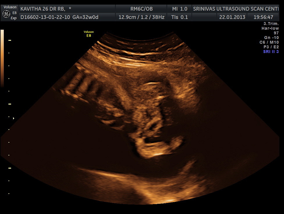
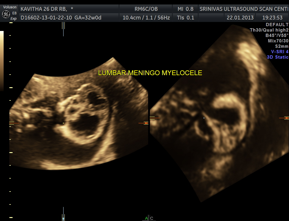
The following picture shows the lumbar meningo myelocele.
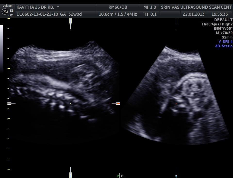
3 d showing 2 views
the following images are 3 d reconstruction.
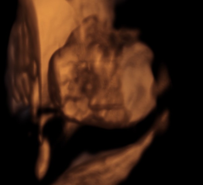
3d reconstruction
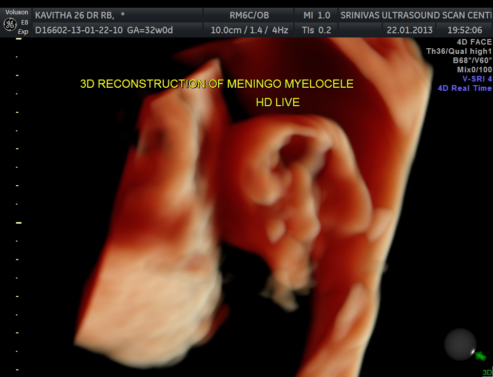
3d live reconstruction


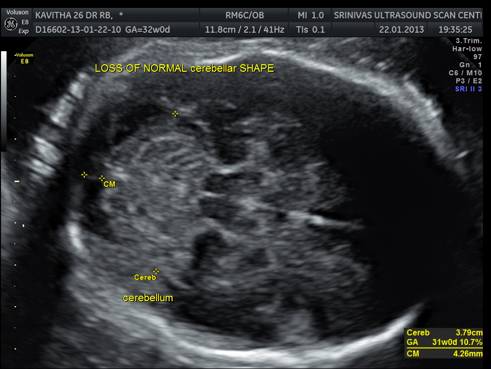
the conventional markers are not very helpful – lemon sign not made out

lemon sign not made out as the GA is around 32 weeks
banana sign is not so clear; but cerebellar shape is changed – boomerang
BPD and HC less than 2.3%tile is a subtle marker to look at the spine closely
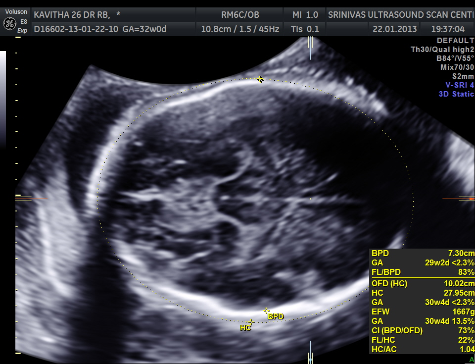
BPD and HC are <2.3%tile and this could be an important marker to look carefully at the spine
COMMENT :
This again illustrates the need to pick up open neural tubal defects earlier , when the lemon and banana sign are much easier to appreciate .
Appreciation of internal translucency in the 1st trimester needs a learning curve , in which all obstetrics practicing sonologists should invest .
Microcephaly could be an important marker in the late 2nd and 3rd trimester scans.

Good thought provoking images and ideas ,we should try to pick up neural tube defect as early as possible .As in India after 20 weeks termination is not allowed
LikeLike
Very valid legal point
LikeLike