25 Year old lady was referred for anomaly scan.
She was gravida 2 , para 1 , miscarriages nil , live 1 – with history of consanguinity. Clinically the uterus size was large for dates ( 27 weeks gestation ) .
Ultrasound showed obvious polyhydramnios.

The following pictures show the anomaly.

the atretic segment is seen in the following picture

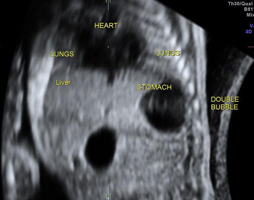
the following image shows the double bubble in a coronal 3d reconstructed image

Few points of interest are given below :
1.the normal duodenum can only occasionally be demonstrated.
2. dilatation of the duodenum as large as or larger than the stomach suggests duodenal atresia or stenosis
3. should not be mistaken for a dilated gall bladder ; demonstrating the continuity between the two bubbles prevents the mistake.
4. the diagnosis is usually made in late 2nd trimester or 3rd trimester and is usually associated with polyhydramnios
5.this can be an isolated anomaly or associated with VATER ( Vertebral,Anorectal,Tracheo Esophageal,Renal /Radial anomalies
6. 30 % association with Down’s syndrome is found
7 If isolated , the post natal outlook is good.

Any follow-up?
LikeLike
Lost for follow up
LikeLike